Conditions · Gastroenterology and hepatology
Choledochal cyst !
A choledochal cyst is an inborn widening of the bile ducts. It can cause jaundice, pain and infections, and carries a risk of bile-duct cancer.
Updated 2026-11-01
Structures involved
Tap one to see what it is and open it in 3D.
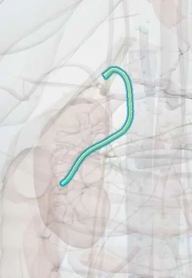
- Common bile duct
- Common hepatic duct
- Gallbladder
- Cystic duct
- Ampulla of Vater
- Pancreatic duct
- Liver
- Duodenum
- Pancreas
Why it happens
- Precancerous conditions: when chronic inflammation, metaplasia or hyperplasia raise the risk
- Exocrine pancreatic secretion
- Bile and the enterohepatic circulation
- Gallbladder and bile ducts
What it is
A congenital cystic dilatation of the extrahepatic and/or intrahepatic bile ducts, classified by the Todani system. Most are found in childhood, but about a quarter are first diagnosed in adults.
Who gets it
Rare in Western countries (about 1 in 100,000–150,000 live births) and much more common in East Asia, particularly Japan. Females are affected three to four times more often than males. Many are now detected on prenatal or routine ultrasound.
Causes
- Anomalous pancreaticobiliary junction with a long common channel outside the sphincter of Oddi, allowing pancreatic juice to reflux into the bile duct (Babbitt theory)
- Distal bile-duct narrowing or obstruction in fetal life
- Possible reduced nerve cells (ganglion cells) in the distal duct
- Type V (Caroli disease) is linked to an inherited ductal plate malformation
Risk factors
- Female sex
- East Asian ancestry
- Anomalous pancreaticobiliary junction
Pathophysiology
When the pancreatic and bile ducts join too early, pancreatic enzymes reflux into the bile duct, inflame and weaken its wall and raise pressure, so the duct dilates. Bile stagnates in the cyst, favouring stones, sludge and bacterial infection. Chronic inflammation and reflux cause metaplasia and dysplasia of the lining, which explains the high lifetime risk of cholangiocarcinoma and gallbladder cancer.
Classification
- Type I: fusiform or cystic dilatation of the extrahepatic duct (most common, about 50–80%), subtypes Ia, Ib, Ic
- Type II: saccular diverticulum of the common bile duct
- Type III: choledochocele, a dilatation of the intraduodenal distal duct
- Type IVa: multiple cysts, intra- and extrahepatic; Type IVb: multiple extrahepatic cysts only
- Type V: multiple intrahepatic cysts only (Caroli disease; Caroli syndrome if hepatic fibrosis is present)
Symptoms and signs
- Classic triad of right upper quadrant pain, jaundice and a palpable mass, present in only a minority (more often in children)
- Intermittent abdominal pain, often in adults
- Jaundice and pale stools in infants
- Nausea and vomiting
- Fever with jaundice during episodes of cholangitis
- Many are found incidentally without symptoms
Diagnosis
- Clinical
- Suspected in a child or young woman with recurrent right upper quadrant pain, jaundice, cholangitis or unexplained pancreatitis, or when a cyst is seen on prenatal ultrasound.
- Tests
- Liver profile showing a cholestatic pattern (raised bilirubin, alkaline phosphatase and GGT) during episodes; amylase or lipase in pancreatitis; blood cultures in cholangitis; tumour markers if malignancy is suspected.
- Imaging
- Abdominal ultrasound is the first test. Magnetic resonance cholangiopancreatography (MRCP) is the preferred test to define the type and the pancreaticobiliary junction; CT helps surgical planning; ERCP is reserved for therapy, since it carries a higher risk of cholangitis and pancreatitis.
Differential diagnosis
- Biliary atresia (in infants)
- Choledocholithiasis with secondary duct dilatation
- Pancreatic pseudocyst
- Hepatic cysts and polycystic liver disease
- Cholangiocarcinoma or pancreatic head tumour causing upstream dilatation
- Duodenal duplication cyst
Goal of treatment
Control acute complications, then remove the cyst completely and restore bile drainage, to stop recurrent infection and greatly reduce the cancer risk.
Treatment
- Acute
- Cholangitis or pancreatitis: fluids, intravenous antibiotics and biliary drainage when needed before definitive surgery.
- Surgical
- Complete excision of the extrahepatic cyst with cholecystectomy and Roux-en-Y hepaticojejunostomy is the standard for types I and IV; type II is excised as a diverticulum. Internal drainage of the cyst into the bowel (cystenterostomy) is no longer used because the cancer risk persists.
- Procedure
- Type III (choledochocele) is often treated endoscopically with sphincterotomy.
- Surgical
- Types IVa and V: liver resection for disease limited to one lobe, or liver transplantation for diffuse disease with complications.
- Follow-up
- Long-term follow-up with liver tests and ultrasound or MRCP, because stricture, stones and cancer can occur even after excision.
Complications
- Recurrent acute cholangitis
- Bile-duct stones and sludge
- Acute pancreatitis
- Cholangiocarcinoma and gallbladder cancer, with risk rising with age
- Secondary biliary cirrhosis and portal hypertension
- Cyst rupture with bile peritonitis
- Liver abscess
Prognosis
Excellent after complete excision in childhood. Risk of malignancy rises with age and is highest after incomplete excision or older drainage operations, so lifelong follow-up is advised; diffuse intrahepatic disease has a less favourable course.
Test yourself
Think of the answer, then tap the question to see it.
Which anatomical anomaly is thought to cause most choledochal cysts?
An anomalous pancreaticobiliary junction with a long common channel, allowing pancreatic juice to reflux into the bile duct.
What is the classic triad of a choledochal cyst, and how common is it?
Right upper quadrant pain, jaundice and a palpable mass; it is present in only a minority of patients.
Which Todani type is the most common, and which type is Caroli disease?
Type I (extrahepatic fusiform or cystic dilatation) is the most common; type V is Caroli disease.
What is the standard surgery for a type I choledochal cyst?
Complete cyst excision with cholecystectomy and Roux-en-Y hepaticojejunostomy.
Which cancer is the main long-term risk of a choledochal cyst?
Cholangiocarcinoma (bile-duct cancer), and also gallbladder cancer.
Related conditions
- Acute cholangitis
- Choledocholithiasis
- Acute pancreatitis
- Cholangiocarcinoma (bile duct cancer)
- Biliary atresia
- Cirrhosis
- Gallstones (cholelithiasis)
Educational information, not medical advice. For any symptom, see a health professional.
Sources: Choledochal Cysts: A Review of Literature (Saudi Journal of Gastroenterology, 2012).
Spotted a mistake?We are always improving for the community. Tell us what is wrong and our team will review it.Report a mistake
Representative model, for educational purposes only.