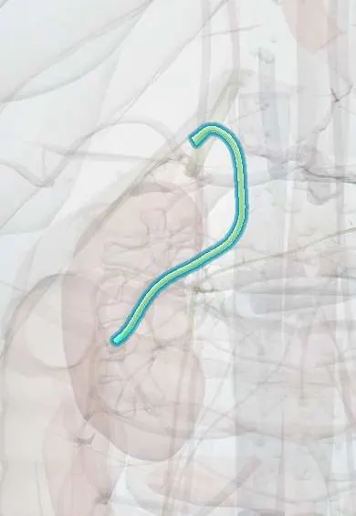
Choledocholithiasis !
Choledocholithiasis means stones in the common bile duct, the tube that carries bile to the gut; they can block it and cause jaundice or infection.
Updated 2026-10-29
Structures involved
Tap one to see what it is and open it in 3D.
- Common bile duct
- Ampulla of Vater
- Common hepatic duct
- Gallbladder
- Cystic duct
- Pancreatic duct
- Duodenum
- Liver
In biochemistry
- Liver function tests: reading the pattern
- Alkaline phosphatase (ALP) as a lab test
- Gamma-glutamyl transferase (GGT) as a lab test
In the clinical exam
In physiology
Why it happens
What it is
Presence of stones in the common bile duct. Most come from the gallbladder (secondary); fewer form in the duct itself (primary).
Who gets it
Found in a significant minority of people with symptomatic gallstones, more often with age. Because gallstones are common in Colombia, it is a frequent reason for ERCP.
Causes
- Gallbladder stones that pass through the cystic duct (secondary, most common)
- Primary duct stones, usually brown pigment, from bile stasis and infection
- Duct strictures, dilatation or parasites (for example Ascaris) that promote stone formation
Risk factors
- Gallbladder stones
- Older age
- Previous cholecystectomy (retained or recurrent stones)
- Biliary strictures or periampullary diverticula
- Biliary parasitic infection
Pathophysiology
A stone in the common bile duct blocks bile flow into the duodenum. Bile backs up, bilirubin and liver enzymes rise and the duct dilates. Stagnant bile can become infected (cholangitis), and a stone at the ampulla can also block the pancreatic duct (pancreatitis).
Classification
- Secondary (from the gallbladder) or primary (formed in the duct)
- Asymptomatic or symptomatic
- Risk of a duct stone graded low, intermediate or high (ASGE criteria); criteria vary between guidelines
Symptoms and signs
- Biliary-type pain in the right upper quadrant or epigastrium
- Jaundice that may come and go
- Dark urine and pale stools
- Itching (pruritus)
- Nausea and vomiting; some stones are found with no symptoms
Diagnosis
- Clinical
- Suspected in a patient with gallstones and jaundice or abnormal liver tests. Risk is graded from clinical, lab and ultrasound findings to choose the next test.
- Tests
- Liver tests with a cholestatic pattern: raised bilirubin, alkaline phosphatase and gamma-glutamyl transferase; transaminases may rise early. Lipase if pancreatitis is suspected.
- Imaging
- Ultrasound shows a dilated common bile duct and sometimes the stone. MRCP or endoscopic ultrasound confirm it in intermediate risk; ERCP is both diagnostic and therapeutic in high risk.
Differential diagnosis
- Pancreatic head or periampullary cancer
- Cholangiocarcinoma
- Benign biliary stricture
- Mirizzi syndrome
- Viral or drug-induced hepatitis
- Primary sclerosing cholangitis
Goal of treatment
Clear the bile duct, prevent cholangitis and pancreatitis, and remove the gallbladder to stop new stones.
Treatment
- Acute
- Supportive care: fluids, analgesia, and antibiotics if there are signs of infection.
- Procedure
- ERCP with sphincterotomy and stone extraction is the main treatment; large stones may need lithotripsy or a temporary stent.
- Surgical
- Laparoscopic cholecystectomy, usually during the same admission after the duct is cleared; some surgeons explore the duct during the operation.
- Procedure
- When ERCP fails or is not possible: percutaneous transhepatic drainage or surgical bile duct exploration.
Complications
- Acute cholangitis
- Acute pancreatitis
- Secondary biliary cirrhosis with long-standing obstruction
- Liver abscess
- Complications of ERCP: pancreatitis, bleeding, perforation
Prognosis
Very good when the duct is cleared and the gallbladder removed. Untreated obstruction can lead to cholangitis, pancreatitis or chronic liver damage.
Prevention
- Cholecystectomy in people with symptomatic gallstones reduces later duct stones
Test yourself
Think of the answer, then tap the question to see it.
What lab pattern suggests a stone in the common bile duct?
A cholestatic pattern: raised bilirubin, alkaline phosphatase and gamma-glutamyl transferase, sometimes with an early rise in transaminases.
Which test both confirms and treats choledocholithiasis?
ERCP, with sphincterotomy and stone extraction.
What non-invasive test confirms a bile duct stone in intermediate-risk patients?
MRCP (magnetic resonance cholangiopancreatography); endoscopic ultrasound is an alternative.
Which two serious complications can a common bile duct stone cause?
Acute cholangitis (infection of obstructed bile) and acute pancreatitis (blockage at the ampulla).
Related conditions
- Gallstones (cholelithiasis)
- Acute cholangitis
- Acute pancreatitis
- Acute cholecystitis
- Pancreatic cancer
- Gallbladder cancer
Educational information, not medical advice. For any symptom, see a health professional.
Sources: Choledocholithiasis (StatPearls (NCBI Bookshelf), 2026).
Spotted a mistake?We are always improving for the community. Tell us what is wrong and our team will review it.Report a mistake
Representative model, for educational purposes only.