Conditions · Hematology and oncology
Essential thrombocythaemia and primary myelofibrosis !
Two related bone marrow cancers that grow slowly: one makes too many platelets, the other scars the marrow so the spleen takes over making blood.
Updated 2026-11-01
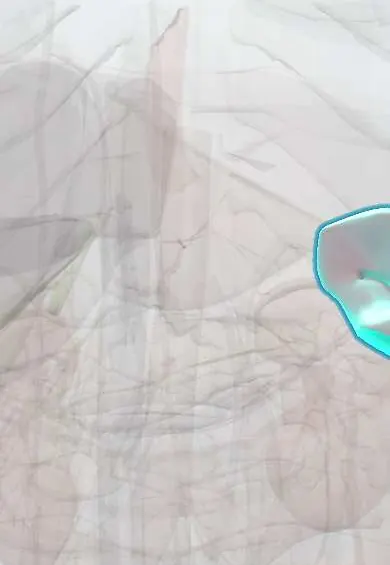
Structures involved
Tap one to see what it is and open it in 3D.
Why it happens
- Oncogenes: RAS, MYC, HER2, BCR-ABL and the table to know
- Fibrosis in organs: TGF-β and the myofibroblast
- Hypercoagulable states: inherited and acquired
- Hematopoiesis
- JAK–STAT pathway
What it is
Two Philadelphia-negative myeloproliferative neoplasms caused by acquired mutations in a blood stem cell. In essential thrombocythaemia (ET) the marrow overproduces platelets; in primary myelofibrosis (PMF) abnormal megakaryocytes drive scarring of the marrow, so blood is made in the spleen and liver instead.
Who gets it
Both are rare and diseases of older adults, usually diagnosed after age 50–60; ET is somewhat more frequent in women and is often found by chance on a routine blood count, while PMF is the least common and most severe of the classic myeloproliferative neoplasms. Latin American registries describe the same mutation pattern as elsewhere; in Colombia, access to molecular testing and newer targeted drugs can delay diagnosis and treatment outside large centres.
Causes
- Acquired driver mutations in haematopoietic stem cells: JAK2 V617F (about half of cases), CALR or MPL
- A minority are 'triple negative', with none of the three driver mutations
- Myelofibrosis can also develop secondarily from long-standing ET or polycythaemia vera (post-ET or post-PV myelofibrosis)
- Fibrosis results from growth factors released by abnormal megakaryocytes, not from an infection or toxin
Risk factors
- Age over 60
- Female sex (ET)
- Previous ET or polycythaemia vera (for secondary myelofibrosis)
- For thrombosis in ET: age over 60, previous thrombosis, JAK2 mutation and cardiovascular risk factors (smoking, hypertension, diabetes)
Pathophysiology
Driver mutations switch on the JAK-STAT signalling pathway without the normal signal from thrombopoietin, so megakaryocytes and platelets multiply. Abnormal platelets and activated white cells promote clotting in arteries and veins, yet very high platelet counts can also cause bleeding by absorbing von Willebrand factor (acquired von Willebrand syndrome). In myelofibrosis, cytokines from abnormal megakaryocytes stimulate fibroblasts to lay down reticulin and collagen; the scarred marrow fails, stem cells move to the spleen and liver (extramedullary haematopoiesis) and inflammatory cytokines cause fatigue, fever, weight loss and night sweats.
Classification
- Essential thrombocythaemia: low-risk or high-risk for thrombosis (age, prior thrombosis, JAK2 status)
- Primary myelofibrosis: prefibrotic/early stage and overt fibrotic stage
- Secondary myelofibrosis: post-ET or post-polycythaemia vera
- Prognostic scores for myelofibrosis (such as IPSS/DIPSS) based on age, symptoms, haemoglobin, white count and blasts
Symptoms and signs
- ET: often no symptoms, found on a routine blood count
- ET: headache, dizziness, visual disturbance, and burning, red painful hands or feet (erythromelalgia)
- Clots in arteries or veins, or bruising and mucosal bleeding when platelets are very high
- PMF: tiredness and breathlessness from anaemia
- PMF: left upper abdominal fullness, early satiety or pain from a very large spleen
- PMF: fever, night sweats, weight loss and itching
Diagnosis
- Clinical
- Suspect ET in a persistently raised platelet count with no reactive cause, and PMF in an older adult with anaemia, constitutional symptoms and a large spleen. Rule out reactive thrombocytosis first (iron deficiency, infection, inflammation, cancer, post-splenectomy).
- Tests
- Full blood count and blood smear: ET shows a sustained high platelet count with large platelets; PMF shows teardrop red cells (dacrocytes) and a leukoerythroblastic picture (immature white and red cells). Mutation testing for JAK2, CALR and MPL, and BCR-ABL to exclude chronic myeloid leukaemia. Bone marrow biopsy is essential: enlarged mature megakaryocytes in ET; megakaryocyte atypia with reticulin or collagen fibrosis in PMF, where aspiration is often a 'dry tap'. Ferritin, LDH and von Willebrand studies when platelets are very high.
- Imaging
- Abdominal ultrasound to measure the spleen and liver and to look for portal or hepatic vein thrombosis; Doppler or CT when splanchnic thrombosis is suspected.
Differential diagnosis
- Reactive (secondary) thrombocytosis from iron deficiency, infection, inflammation, cancer or splenectomy
- Polycythaemia vera
- Chronic myeloid leukaemia (BCR-ABL positive)
- Myelodysplastic syndrome, including overlap forms
- Secondary marrow fibrosis from metastatic cancer, tuberculosis or other granulomatous infection
Goal of treatment
In ET, prevent thrombosis and bleeding according to risk; in PMF, control anaemia, spleen size and symptoms, and offer cure by stem cell transplant to suitable patients.
Treatment
- Medical
- ET: low-dose aspirin for most patients without bleeding risk; cytoreduction for high-risk patients, with hydroxyurea first line and interferon alfa (preferred in pregnancy and younger patients) or anagrelide as alternatives.
- Lifestyle
- Control of cardiovascular risk factors (stopping smoking, treating hypertension, diabetes and dyslipidaemia) to lower clotting risk.
- Medical
- PMF: JAK inhibitors (such as ruxolitinib) to shrink the spleen and relieve symptoms; treatment of anaemia with transfusions, erythropoiesis-stimulating agents or androgens, and iron chelation when transfusions are frequent.
- Procedure
- Allogeneic haematopoietic stem cell transplantation is the only potentially curative treatment for myelofibrosis, reserved for fitter patients with intermediate- or high-risk disease.
- Follow-up
- Regular blood counts and spleen checks; nursing care focuses on teaching the signs of clotting and bleeding, fall and fatigue management, and monitoring of cytopenias during cytoreductive or JAK inhibitor therapy.
Drugs for this condition
Complications
- Arterial and venous thrombosis, including unusual sites such as the portal and hepatic veins
- Bleeding from acquired von Willebrand syndrome at very high platelet counts
- Progression of ET to myelofibrosis
- Transformation to acute myeloid leukaemia
- In PMF: massive splenomegaly, splenic infarction, portal hypertension and transfusion iron overload
Prognosis
Essential thrombocythaemia has a near-normal life expectancy when thrombotic risk is managed. Primary myelofibrosis has the worst prognosis of the classic myeloproliferative neoplasms, with median survival ranging from many years in low-risk patients to only a few years in high-risk disease; transplant offers the only chance of cure.
Test yourself
Think of the answer, then tap the question to see it.
Which three driver mutations are tested for in essential thrombocythaemia and primary myelofibrosis?
JAK2 V617F, CALR and MPL.
What blood smear finding is classic for primary myelofibrosis?
Teardrop red cells (dacrocytes) with a leukoerythroblastic picture.
Why is bone marrow aspiration often a 'dry tap' in myelofibrosis?
Reticulin and collagen fibrosis trap the cells, so a core biopsy is needed.
What is the first-line cytoreductive drug for high-risk essential thrombocythaemia?
Hydroxyurea, usually combined with low-dose aspirin.
Which treatment is the only potentially curative option for primary myelofibrosis?
Allogeneic haematopoietic stem cell transplantation.
Related conditions
- Polycythaemia vera
- Chronic myeloid leukaemia
- Myelodysplastic syndromes
- Acute leukaemia (AML and ALL)
- Iron deficiency anaemia
- Budd-Chiari syndrome
- Portal vein thrombosis
- Deep vein thrombosis
- Ischaemic stroke
- Von Willebrand disease
- Gout
Educational information, not medical advice. For any symptom, see a health professional.
Sources: Essential thrombocythemia (MedlinePlus (U.S. National Library of Medicine), 2025); Myelofibrosis (MedlinePlus (U.S. National Library of Medicine), 2026).
Spotted a mistake?We are always improving for the community. Tell us what is wrong and our team will review it.Report a mistake
Representative model, for educational purposes only.