Cardiovascular system · Abdomen
Splenic vein !
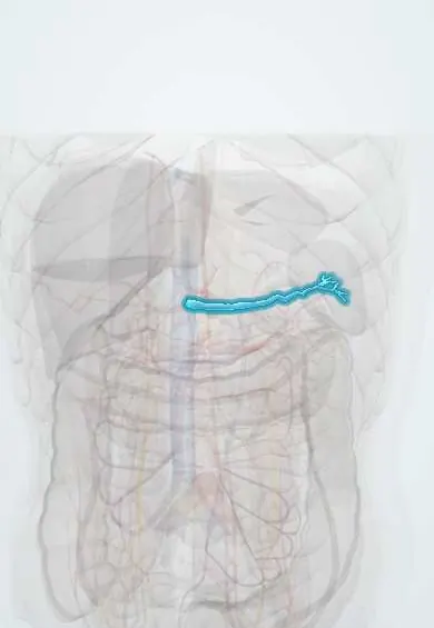
The splenic vein drains blood from the spleen and runs along the back of the pancreas to help form the portal vein.
Opens in the Durgical app, no sign-up needed.
What it is
The vein from the spleen running behind the pancreas to join the portal vein.
What it does
Drains the spleen, pancreas and part of the stomach.
Key facts
- Drains
- Spleen; tributaries: short gastric, left gastroepiploic, pancreatic veins and usually the inferior mesenteric vein
- Into
- Runs in a groove on back of pancreas and joins SMV behind pancreatic neck to form the portal vein
Relations
- Course
- Leaves the splenic hilum, runs in the splenorenal ligament with the splenic artery and the tail of the pancreas, then straight to the right along the back of the pancreas to the neck, where it meets the superior mesenteric vein
- In front
- Body and tail of the pancreas, which partly embed it in a groove
- Behind
- Left kidney and left adrenal gland laterally; near the midline the superior mesenteric artery, with the left renal vein and the abdominal aorta behind it
- Above
- Splenic artery, which runs a more tortuous course along the upper border of the pancreas
How to find it
Not palpable. At surgery it is found on the back of the pancreas by lifting the lower border of the body and tail; it lies in a groove there and receives many short pancreatic veins, which have to be tied one by one to keep the spleen. On ultrasound it is the tubular line just behind the pancreas that ends at the confluence with the superior mesenteric vein.
On imaging
Ultrasound and contrast CT show it as a straight tube behind the pancreas; in pancreatitis or a pancreatic mass, a missing or thin vein with large collaterals around the stomach suggests thrombosis. Endoscopy shows the gastric varices.
Common problems
- Portal hypertension in cirrhosis: blood backs up, causing swollen veins in the esophagus (varices) that can bleed, an enlarged spleen and fluid in the abdomen (ascites).
How it is diagnosed
Ultrasound with Doppler, CT, endoscopy for varices.
Treatment
- Medicines that lower the pressure, banding of varices, a shunt (TIPS), liver transplant.
Clinical notes
- In left-sided portal hypertension removing the spleen cures the gastric varices, because it removes the inflow that feeds them
- Distal splenorenal shunt: in portal hypertension the splenic vein can be joined to the left renal vein to decompress varices
- Its close bond with the pancreas makes it the vessel most at risk in distal pancreatectomy and during spleen-preserving surgery
Anatomical variants
- The inferior mesenteric vein often ends in the splenic vein; it may instead join the superior mesenteric vein or the confluence of the two (texts differ on which is commonest)
- The left gastric (coronary) vein may drain into the splenic vein instead of the portal vein or the confluence, which matters in surgery for portal hypertension
Related conditions
Test yourself
Think of the answer, then tap the question to see it.
What is the course of the splenic vein?
From the splenic hilum through the splenorenal ligament, then straight to the right along the back of the pancreas to its neck, where it joins the superior mesenteric vein.
What does splenic vein thrombosis cause?
Left-sided (sinistral) portal hypertension: isolated gastric fundal varices through the short gastric veins, with a normal liver; mostly from pancreatitis or a pancreatic tumour.
Why does splenectomy cure the gastric varices of left-sided portal hypertension?
It removes the inflow from the spleen that feeds them.
Which veins drain into the splenic vein?
The short gastric, left gastro-omental and pancreatic veins, and usually the inferior mesenteric vein.
Educational information, not medical advice. For any symptom, see a health professional.
Its names
- Official Latin (TA2)
- vena splenica TA2 5123
- Official Spanish (DeCS)
- Vena esplénica DeCS D013162
- English (TA2)
- Splenic vein
- Also called
- lienal vein
Related
Sources: Terminologia Anatomica (TA2); DeCS; Anatomy, Abdomen and Pelvis, Portal Venous System (Hepatic Portal System); 20.5 Circulatory Pathways (OpenStax (Rice University), 2022) · adapted, CC BY 4.0.
Spotted a mistake?We are always improving for the community. Tell us what is wrong and our team will review it.Report a mistake
Representative model, for educational purposes only.