Conditions · Systemic conditions
Gout !
Gout is arthritis caused by uric acid crystals building up in joints; it causes sudden, very painful attacks, classically in the big toe.
Updated 2026-10-29
Structures involved
Tap one to see what it is and open it in 3D.
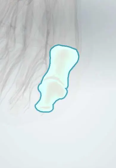
- Big toe
- MTP ligaments and plantar plate
- Joint capsule
- First metatarsal
- Toe joint capsules
- Prepatellar bursa
- Kidneys
- Ureters
In biochemistry
- De novo purine synthesis: Step 1 · PRPP synthetase
- Purine degradation and salvage: Step 5 · Xanthine oxidase
- Glucose-6-phosphatase
- Ribose 5-phosphate
- Fructose
- PRPP synthetase
- Glutamine-PRPP amidotransferase
- Hypoxanthine-guanine phosphoribosyltransferase (HGPRT)
- Xanthine oxidase
- PRPP (phosphoribosyl pyrophosphate)
- Uric acid
- Drugs acting on nucleotide metabolism
- Types of enzyme inhibition
In histology
In the clinical exam
In physiology
Why it happens
- Mediators of inflammation: the table to know
- When leukocytes injure tissue: proteases, oxidants and NETs
- Other forms of cell death: necroptosis, pyroptosis, ferroptosis
- Inflammation: the cardinal signs and its five steps
- Arachidonic acid metabolites in inflammation, and the drugs that block them
- Heavy metals: lead, mercury, arsenic and cadmium
Show 1 more
What it is
An inflammatory arthritis caused by monosodium urate crystals depositing in and around joints when uric acid stays high for a long time. It causes sudden, very painful flares, classically in the big toe, and can lead to tophi and kidney stones.
Who gets it
The most common inflammatory arthritis in adult men; in women it appears mostly after menopause. It is linked to obesity, metabolic syndrome, hypertension and kidney disease, which are rising in Latin America.
Causes
- Hyperuricaemia from reduced kidney excretion of uric acid (most cases)
- Overproduction of urate: high cell turnover (myeloproliferative disorders, tumour lysis), rare enzyme defects
- Diet rich in purines (red meat, organ meats, seafood), alcohol (especially beer) and fructose-sweetened drinks
- Drugs: thiazide and loop diuretics, low-dose aspirin, cyclosporine
Risk factors
- Male sex and older age; women after menopause
- Obesity and metabolic syndrome
- Hypertension and chronic kidney disease
- Alcohol and sugary drinks
- Diuretic use
- Family history
Pathophysiology
Above about 6.8 mg/dL urate exceeds its solubility and forms needle-shaped crystals, favoured by cooler, acidic or damaged joints such as the first metatarsophalangeal joint. Macrophages phagocytose crystals and activate the NLRP3 inflammasome, releasing IL-1β and recruiting neutrophils. Chronic deposits form tophi that erode bone.
Classification
- Asymptomatic hyperuricaemia (not gout)
- Acute gout flare
- Intercritical (symptom-free) periods
- Chronic tophaceous gout
Symptoms and signs
- Sudden, severe joint pain that peaks within 24 hours, often starting at night
- Podagra: red, hot, swollen first metatarsophalangeal joint (big toe)
- Other joints: midfoot, ankle, knee, wrist, fingers, olecranon bursa
- Exquisite tenderness; even a bed sheet hurts
- Fever possible in large or multiple flares
- Tophi: firm, chalky nodules on the ear, fingers, elbows or Achilles tendon
Diagnosis
- Clinical
- A typical flare in the big toe with a known high urate suggests gout, but crystal identification is the gold standard; a clinical rule may be used where aspiration is not possible.
- Tests
- Joint aspiration: needle-shaped, negatively birefringent urate crystals under polarised light, with Gram stain and culture to exclude infection. Serum urate can be normal during a flare and is best rechecked a few weeks later; kidney function, glucose and lipids.
- Imaging
- Ultrasound shows the double-contour sign and tophi; dual-energy CT detects urate deposits. X-rays in chronic gout show punched-out erosions with overhanging edges.
Differential diagnosis
- Septic arthritis
- Calcium pyrophosphate deposition (pseudogout)
- Cellulitis
- Reactive or psoriatic arthritis
- Rheumatoid arthritis (in chronic polyarticular gout)
- Fracture or trauma
Goal of treatment
Relieve the flare quickly and, in recurrent or tophaceous gout, lower serum urate long term so crystals dissolve and flares, tophi and joint damage stop.
Treatment
- Acute
- Flare: NSAIDs, colchicine or glucocorticoids (oral or intra-articular), started as early as possible and chosen according to kidney function and other illnesses; rest and ice.
- Medical
- Urate-lowering therapy (xanthine oxidase inhibitors first line; uricosurics or uricase agents in selected cases) for recurrent flares, tophi, urate stones or joint damage, aiming for a target urate.
- Medical
- Flare prophylaxis with low-intensity anti-inflammatory therapy during the first months of urate-lowering therapy.
- Lifestyle
- Weight loss, less alcohol and sugary drinks, fewer purine-rich foods, and review of diuretics where possible.
- Follow-up
- Serum urate checks to adjust urate-lowering therapy to target, and kidney function.
Drugs for this condition
Complications
- Chronic tophaceous gout with joint destruction
- Uric acid kidney stones
- Chronic kidney disease (often both cause and effect)
- Tophus ulceration or infection
- Associated cardiovascular disease
Prognosis
Flares settle in days to two weeks with treatment. With urate kept at target, flares stop and tophi shrink over months to years; without it, flares become more frequent and polyarticular.
Prevention
- Healthy weight and limiting alcohol and fructose drinks
- Urate-lowering therapy continued long term once indicated
- Managing associated hypertension, diabetes and kidney disease
Test yourself
Think of the answer, then tap the question to see it.
What do urate crystals look like under polarised light?
Needle-shaped and strongly negatively birefringent (yellow when parallel to the compensator axis); pseudogout crystals are rhomboid and weakly positive.
What is podagra?
A gout flare in the first metatarsophalangeal joint of the big toe: sudden pain, redness, heat and swelling; it is the classic first presentation.
Why can serum uric acid be normal during a gout flare?
Inflammation increases urate excretion, so levels may fall during a flare; a normal value does not exclude gout and is rechecked later.
What is the main difference between flare treatment and urate-lowering therapy?
Flare treatment (NSAIDs, colchicine, steroids) controls inflammation; urate-lowering therapy (for example xanthine oxidase inhibitors) dissolves crystals and prevents future flares.
What are tophi?
Collections of urate crystals surrounded by inflammatory tissue, felt as firm nodules (ear, fingers, elbow); they mark chronic, poorly controlled gout.
Related conditions
- Gout of the Knee
- Pseudogout (CPPD) of the Knee
- Septic Arthritis of the Knee
- Rheumatoid arthritis
- Diabetic foot
- Cellulitis of the leg
- Hallux valgus (bunion)
- Olecranon bursitis
Educational information, not medical advice. For any symptom, see a health professional.
Sources: Gout (StatPearls (NCBI Bookshelf), 2026).
Spotted a mistake?We are always improving for the community. Tell us what is wrong and our team will review it.Report a mistake
Representative model, for educational purposes only.