Conditions · Nephrology and urology
Fanconi syndrome !
A failure of the kidney's proximal tubule, so glucose, amino acids, phosphate and bicarbonate leak into the urine. Not the same as Fanconi anaemia.
Updated 2026-11-01
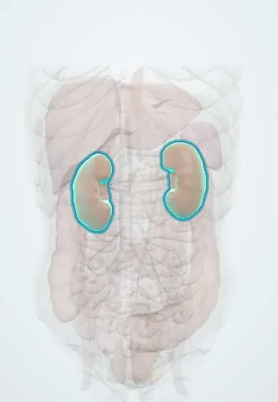
Structures involved
Tap one to see what it is and open it in 3D.
Why it happens
- Heavy metals: lead, mercury, arsenic and cadmium
- Proximal tubule transport
- Glucose transport maximum and renal threshold
- Renal handling of H⁺ and bicarbonate
- Primary and secondary active transport
- Kidney: cortex, medulla and the nephron
What it is
A generalised dysfunction of the proximal convoluted tubule causing urinary loss of substances it normally reabsorbs: glucose (with normal blood glucose), amino acids, phosphate, bicarbonate, urate, potassium and low-molecular-weight proteins. It is distinct from Fanconi anaemia, an inherited bone-marrow failure disorder.
Who gets it
Rare. In children, cystinosis is the most common inherited cause; other inborn errors include tyrosinaemia type 1, galactosaemia, Wilson disease and hereditary fructose intolerance. In adults most cases are acquired, from drugs (tenofovir and older antiretrovirals, ifosfamide, aminoglycosides, valproate), monoclonal light chains (multiple myeloma) or heavy metals, which is relevant where HIV therapy and lead exposure are common.
Causes
- Inherited: cystinosis, tyrosinaemia type 1, galactosaemia, hereditary fructose intolerance, Wilson disease, Dent disease, Lowe syndrome
- Drugs: tenofovir and some older antiretrovirals, ifosfamide and cisplatin, aminoglycosides, valproate, expired tetracyclines
- Paraproteins: light-chain disease and multiple myeloma
- Heavy metals: lead, cadmium, mercury
- After kidney transplant or with some autoimmune kidney disease
Risk factors
- Family history or consanguinity (inherited metabolic disorders)
- Treatment with nephrotoxic antiretrovirals, chemotherapy or anticonvulsants
- Monoclonal gammopathy or myeloma
- Occupational or environmental heavy-metal exposure
Pathophysiology
Proximal tubular cells reabsorb most filtered solutes using sodium-coupled transporters powered by ATP. Toxic metabolites, drugs or light chains injure these cells or their energy supply, so many transporters fail together. Bicarbonate wasting causes proximal (type 2) renal tubular acidosis; phosphate wasting causes rickets or osteomalacia; glucose, amino acid and urate losses appear in the urine; sodium and water loss cause polyuria and volume depletion, and increased distal delivery causes potassium loss.
Classification
- Inherited (primary or secondary to an inborn error of metabolism) versus acquired
- Complete (multiple solutes) versus partial (isolated defects)
Symptoms and signs
- Polyuria, thirst and dehydration
- Children: failure to thrive and poor growth
- Bone pain, bowed legs (rickets) or fractures and osteomalacia in adults
- Muscle weakness from hypokalaemia and hypophosphataemia
- Features of the cause: corneal cystine crystals and photophobia (cystinosis), liver disease (tyrosinaemia, Wilson)
Diagnosis
- Clinical
- Suspected with polyuria, growth failure or rickets in a child, or with metabolic acidosis and hypophosphataemia in an adult taking a nephrotoxic drug.
- Tests
- Normal anion gap metabolic acidosis with hypokalaemia and hypophosphataemia; glucosuria with normal blood glucose; generalised aminoaciduria; phosphaturia (low tubular phosphate reabsorption); low uric acid; low-molecular-weight proteinuria. Targeted tests for the cause: leukocyte cystine, succinylacetone, serum and urine protein electrophoresis with free light chains, copper studies, lead level.
- Imaging
- X-rays show rickets or osteomalacia; kidney ultrasound may show nephrocalcinosis or stones in some causes.
Differential diagnosis
- Isolated proximal (type 2) renal tubular acidosis
- Distal (type 1) renal tubular acidosis
- Diabetes mellitus (glucosuria with high blood glucose)
- Nutritional or vitamin D-dependent rickets
- Bartter or Gitelman syndrome (hypokalaemia with alkalosis rather than acidosis)
Goal of treatment
Replace urinary losses, correct acidosis and protect bone and growth, while treating or removing the cause.
Treatment
- Medical
- Replacement of fluids and electrolytes: alkali (bicarbonate or citrate), potassium and phosphate, with active vitamin D to support bone.
- Medical
- Treat or remove the cause: stop the offending drug, treat myeloma, chelation for heavy metals, diet or targeted therapy for metabolic disorders.
- Medical
- Cystinosis: lifelong cysteamine therapy depletes cell cystine and slows kidney and organ damage; nitisinone for tyrosinaemia type 1.
- Follow-up
- Monitoring of growth, electrolytes, acid-base status, kidney function and bone health.
- Surgical
- Kidney transplant for kidney failure (for example in cystinosis); the graft does not develop the defect.
Complications
- Rickets, growth failure and osteomalacia
- Severe dehydration and electrolyte emergencies
- Chronic kidney disease (especially in cystinosis)
- Kidney stones or nephrocalcinosis in some forms
Prognosis
Depends on the cause. Drug-induced Fanconi syndrome often improves after the drug is stopped. Untreated nephropathic cystinosis leads to kidney failure in childhood; early cysteamine delays it substantially.
Test yourself
Think of the answer, then tap the question to see it.
Which part of the nephron is affected in Fanconi syndrome?
The proximal convoluted tubule, with generalised failure of reabsorption.
What type of renal tubular acidosis occurs in Fanconi syndrome?
Proximal (type 2) RTA from bicarbonate wasting, a normal anion gap metabolic acidosis.
What is the most common inherited cause of Fanconi syndrome in children?
Cystinosis.
Which commonly tested drug is a cause of acquired Fanconi syndrome?
Tenofovir (also ifosfamide, aminoglycosides, valproate and expired tetracyclines).
How is glucosuria in Fanconi syndrome distinguished from that in diabetes?
In Fanconi syndrome blood glucose is normal; the glucose is lost because the proximal tubule cannot reabsorb it.
Related conditions
- Renal tubular acidosis
- Wilson disease
- Tyrosinaemia type I (hepatorenal)
- Classic galactosaemia
- Multiple myeloma
- Rickets and osteomalacia
- Hypophosphataemia
- Hypokalaemia
- Lead poisoning
- Chronic kidney disease
Educational information, not medical advice. For any symptom, see a health professional.
Sources: Fanconi syndrome: MedlinePlus Medical Encyclopedia (MedlinePlus (U.S. National Library of Medicine), 2024).
Spotted a mistake?We are always improving for the community. Tell us what is wrong and our team will review it.Report a mistake
Representative model, for educational purposes only.