Conditions · Nephrology and urology
Hypophosphataemia !
Low phosphate in the blood. When severe, as in refeeding after starvation, it weakens muscles, including those used to breathe and the heart.
Updated 2026-11-01
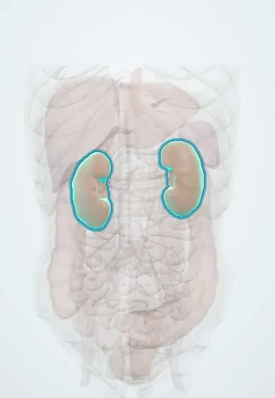
Structures involved
Tap one to see what it is and open it in 3D.
Why it happens
- Phosphate and FGF-23
- Renal handling of calcium, phosphate and magnesium
- Calcium and phosphate
- ATP (adenosine triphosphate)
- Hemoglobin and 2,3-BPG
What it is
A serum phosphate below the normal range (about 2.5 mg/dL in adults). Mild cases are common and silent; severe hypophosphataemia (below about 1 mg/dL) depletes cell energy (ATP) and can cause muscle, respiratory, cardiac and blood-cell failure. Refeeding syndrome is its most important acute cause.
Who gets it
Common in hospitals, especially in intensive care, in people with alcohol use disorder, during treatment of diabetic ketoacidosis, and when malnourished patients are fed again. In Colombia and Latin America, childhood and adult undernutrition and alcohol use make refeeding hypophosphataemia relevant on wards and in nutrition units.
Causes
- Shift into cells: refeeding after starvation, insulin treatment of diabetic ketoacidosis, acute respiratory alkalosis, hungry bone syndrome after parathyroidectomy
- Increased kidney loss: primary or secondary hyperparathyroidism, Fanconi syndrome, inherited phosphate-wasting disorders (FGF23 excess), some drugs
- Reduced intestinal absorption: malnutrition, chronic diarrhoea or malabsorption, phosphate-binding antacids, vitamin D deficiency
- Alcohol use disorder, which combines poor intake, losses and shifts
Risk factors
- Prolonged fasting, anorexia nervosa or severe malnutrition before nutrition is restarted
- Chronic alcohol use
- Diabetic ketoacidosis under treatment
- Critical illness, sepsis and mechanical ventilation
- Vitamin D deficiency and hyperparathyroidism
Pathophysiology
Phosphate is needed to make ATP and 2,3-diphosphoglycerate. When a starved person receives carbohydrate, insulin drives glucose, phosphate, potassium and magnesium into cells, so serum phosphate falls sharply. Low ATP impairs contraction of skeletal, respiratory and cardiac muscle and nerve function; low 2,3-DPG shifts the oxygen curve left, reducing oxygen delivery; red cells may haemolyse. Chronic loss demineralises bone (rickets, osteomalacia).
Classification
- Mild: just below normal, usually asymptomatic
- Moderate
- Severe: about 1 mg/dL or lower, with risk of organ dysfunction
- By mechanism: redistribution, renal loss or reduced intestinal absorption
Symptoms and signs
- Often no symptoms in mild cases
- Muscle weakness and myalgia
- Confusion, irritability, paraesthesias or seizures in severe cases
- Breathlessness from respiratory muscle weakness
- Bone pain and deformities in chronic deficiency (osteomalacia, rickets)
Diagnosis
- Clinical
- Suspected in at-risk patients (malnutrition, alcohol, DKA treatment, ICU) with weakness, respiratory difficulty or confusion; the cause is sought in history and medications.
- Tests
- Serum phosphate, with calcium, magnesium and potassium (often also low). Urine phosphate (fractional excretion or 24-hour) separates kidney loss from shifts or poor intake. PTH, vitamin D, and FGF23 in selected cases; CK if rhabdomyolysis is suspected.
- Imaging
- Not needed for the electrolyte disorder; X-rays or bone densitometry when osteomalacia or rickets is suspected.
Differential diagnosis
- Hypokalaemia or hypomagnesaemia causing weakness (often coexist)
- Guillain-Barré syndrome or critical-illness myopathy in weak ICU patients
- Spurious low phosphate (e.g. some drugs or lab interference)
- Primary hyperparathyroidism versus vitamin D deficiency as the cause
Goal of treatment
Correct the phosphate deficit safely, treat the cause, and prevent refeeding syndrome in people at risk.
Treatment
- Medical
- Oral phosphate replacement for mild to moderate, asymptomatic cases, together with the other electrolytes that are low.
- Acute
- Intravenous phosphate for severe or symptomatic hypophosphataemia, given slowly with frequent checks to avoid hypocalcaemia and precipitation.
- Medical
- Treat the cause: vitamin D replacement, management of hyperparathyroidism, stopping phosphate-binding antacids, managing alcohol use.
- Follow-up
- Refeeding prevention: identify at-risk patients, start nutrition slowly, give thiamine, and check phosphate, potassium and magnesium daily during the first days.
- Follow-up
- Nursing monitoring of muscle strength, respiratory effort, heart rhythm and fluid balance during correction.
Complications
- Respiratory failure and difficult ventilator weaning
- Heart failure and arrhythmias
- Rhabdomyolysis
- Haemolysis and impaired white-cell and platelet function
- Osteomalacia and rickets in chronic deficiency
Prognosis
Usually corrects well when the cause is treated. Severe hypophosphataemia in critically ill or refeeding patients is linked to higher mortality, largely through cardiac and respiratory failure, which is why prevention and monitoring matter.
Prevention
- Screening malnourished patients for refeeding risk before feeding
- Gradual reintroduction of nutrition with electrolyte monitoring
- Phosphate monitoring during DKA treatment and in the ICU
Test yourself
Think of the answer, then tap the question to see it.
What is the classic setting for severe acute hypophosphataemia?
Refeeding a starved or severely malnourished patient, when insulin drives phosphate into cells.
Why does severe hypophosphataemia cause muscle and respiratory weakness?
Phosphate is needed to make ATP; depletion impairs contraction of skeletal and respiratory muscles.
Which red-cell compound falls with hypophosphataemia, reducing oxygen delivery?
2,3-diphosphoglycerate (2,3-DPG), shifting the oxygen-haemoglobin curve to the left.
Which urine test separates renal phosphate wasting from other causes?
Urine phosphate, as fractional excretion or a 24-hour collection: high despite low serum phosphate means renal loss.
Related conditions
- Refeeding syndrome
- Rickets and osteomalacia
- Primary hyperparathyroidism
- Alcohol use disorder
- Diabetic ketoacidosis
- Anorexia nervosa
- Fanconi syndrome
- Rhabdomyolysis
- Hypomagnesaemia
- Hypokalaemia
Educational information, not medical advice. For any symptom, see a health professional.
Sources: Phosphorus blood test: MedlinePlus Medical Encyclopedia (MedlinePlus (U.S. National Library of Medicine), 2024).
Spotted a mistake?We are always improving for the community. Tell us what is wrong and our team will review it.Report a mistake
Representative model, for educational purposes only.