Conditions · Pediatrics and neonatology
Hemolytic uremic syndrome (HUS) !
HUS is a serious complication, mostly in young children after bloody diarrhoea, in which tiny clots damage the kidneys, destroy red cells and use up platelets.
Updated 2026-11-01
Structures involved
Tap one to see what it is and open it in 3D.
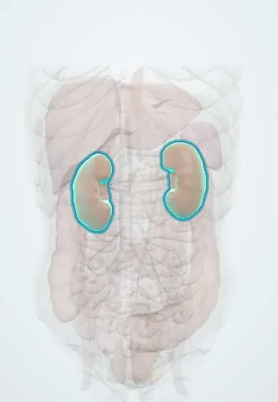
- Kidneys
- Descending colon
- Vessels inside the kidney
- Cecum
- Ascending colon
- Transverse colon
- Sigmoid colon
- Pancreas
Microorganisms that cause it
Why it happens
- Thrombosis and Virchow's triad
- Physiology of acute and chronic kidney failure
- Ribosome (and the antibiotics that target it)
- Glomerulus and the filtration barrier: podocytes, mesangium, JGA
What it is
A thrombotic microangiopathy defined by the triad of microangiopathic haemolytic anaemia, thrombocytopenia and acute kidney injury. The typical form follows bloody diarrhoea caused by Shiga toxin-producing Escherichia coli (STEC) and is the leading cause of acute kidney failure in young children.
Who gets it
Mostly affects children under 5 years. Up to about 1 in 10 people with STEC infection develop HUS. Argentina has one of the highest childhood rates in the world and treats it as a notifiable public-health problem; in Colombia and the rest of Latin America it is less often reported, but STEC from undercooked beef, raw milk and unsafe water is a recognised risk. Atypical (complement-mediated) HUS is rare and can occur at any age.
Causes
- Typical (STEC-HUS, about 90% of children): Shiga toxin-producing E. coli, classically serotype O157:H7; less often Shigella dysenteriae type 1
- Pneumococcal HUS: invasive Streptococcus pneumoniae infection (pneumonia with empyema, meningitis)
- Atypical HUS: genetic or acquired dysregulation of the alternative complement pathway (e.g. factor H, factor I or MCP variants, anti-factor H antibodies)
- Secondary causes: some drugs (calcineurin inhibitors, some chemotherapy agents), transplantation, pregnancy, malignant hypertension, cobalamin C defect
Risk factors
- Age under 5 years (and older adults) with STEC infection
- Eating undercooked ground beef, raw (unpasteurised) milk or dairy, contaminated raw vegetables or sprouts
- Drinking or swimming in unsafe water; contact with farm animals
- Person-to-person spread in households and day-care centres
- Use of antibiotics or antimotility drugs during STEC diarrhoea, which is associated with a higher risk of HUS
- Family history of HUS (atypical form)
Pathophysiology
STEC colonises the colon and releases Shiga toxin, which crosses into the blood and binds the Gb3 receptor, abundant on glomerular endothelium. The toxin halts protein synthesis and injures endothelial cells, triggering platelet-rich microthrombi in small vessels of the kidney (and sometimes brain, gut and pancreas). Red cells are sheared as they pass the damaged vessels (schistocytes), platelets are consumed, and reduced glomerular blood flow causes acute kidney injury. In atypical HUS, uncontrolled complement activation produces the same endothelial damage without an infection trigger.
Classification
- Typical HUS: STEC-associated, preceded by diarrhoea (D+ HUS)
- Pneumococcal HUS: neuraminidase exposes the Thomsen-Friedenreich antigen; direct Coombs test often positive
- Atypical HUS: complement-mediated, no diarrhoeal prodrome, relapsing course
- Secondary HUS: drugs, transplantation, pregnancy, systemic disease
Symptoms and signs
- Prodrome of abdominal cramps and diarrhoea, often bloody, with little or no fever
- About 5 to 10 days after the diarrhoea starts: sudden pallor, tiredness and irritability
- Reduced urine output or no urine (oliguria or anuria), sometimes dark or bloody urine
- Swelling (oedema) and high blood pressure
- Petechiae or bruising from low platelets, usually without major bleeding
- Mild jaundice; enlarged liver or spleen on examination
Diagnosis
- Clinical
- Suspected in a child with recent bloody diarrhoea who becomes pale, swollen and passes little urine. The examination checks hydration, blood pressure, weight, oedema, petechiae, abdominal signs and neurological status.
- Tests
- Complete blood count: anaemia and platelets below 150,000/µL. Peripheral smear with schistocytes. Haemolysis markers: high LDH and indirect bilirubin, low haptoglobin, raised reticulocytes; direct Coombs test negative (positive in pneumococcal HUS). Coagulation times usually normal, which helps separate it from DIC. Rising creatinine and urea, electrolytes (hyperkalaemia, acidosis), urinalysis with haematuria and proteinuria. Stool culture and testing for Shiga toxin or its genes (PCR). In atypical cases: complement C3, ADAMTS13 activity to exclude TTP, and genetic studies.
- Imaging
- Not required for diagnosis. Kidney ultrasound may show enlarged, echogenic kidneys and rules out obstruction; abdominal ultrasound assesses colitis or intussusception; brain MRI if there are neurological signs.
Differential diagnosis
- Thrombotic thrombocytopenic purpura (ADAMTS13 activity below 10%, more neurological signs, mostly adults)
- Disseminated intravascular coagulation (prolonged coagulation times, low fibrinogen)
- Immune thrombocytopenia or Evans syndrome (no kidney injury; Coombs positive in Evans)
- Acute kidney injury from dehydration in gastroenteritis (no haemolysis or schistocytes)
- Intussusception, inflammatory bowel disease or bacterial dysentery as causes of bloody diarrhoea
- Sepsis, malignant hypertension and systemic lupus erythematosus
Goal of treatment
Support the patient through the acute phase: keep fluid and electrolyte balance, correct severe anaemia, control blood pressure, replace kidney function when needed and detect extrarenal complications early, while avoiding interventions that may worsen the disease.
Treatment
- Acute
- Supportive care in hospital, often in a paediatric unit with access to dialysis. Early isotonic intravenous fluids during STEC diarrhoea may protect the kidneys; once oliguria sets in, fluids are restricted and matched to losses.
- Acute
- Red cell transfusion for severe anaemia. Platelet transfusion is generally avoided unless there is active bleeding or a procedure, because it may add to microthrombi.
- Procedure
- Kidney replacement therapy (peritoneal dialysis or haemodialysis) for anuria, refractory hyperkalaemia, acidosis, fluid overload or uraemia.
- Medical
- Antihypertensive drugs for high blood pressure. In STEC diarrhoea, antibiotics and antimotility agents are generally avoided. Atypical HUS is treated with complement C5 inhibitors (such as eculizumab); plasma exchange is used when TTP or complement disease is suspected and the inhibitor is not available.
- Follow-up
- Nursing priorities: strict intake and output, daily weight, frequent blood pressure, neurological checks, electrolytes and blood counts; contact precautions and hand hygiene to prevent spread to others.
Complications
- Need for acute dialysis (about half of children with typical HUS)
- Chronic kidney disease, proteinuria and hypertension years later
- Seizures, coma or stroke from central nervous system involvement
- Haemorrhagic colitis with bowel necrosis, perforation or later strictures
- Pancreatitis and diabetes mellitus; heart involvement (myocardial dysfunction)
- Fluid overload, pulmonary oedema and life-threatening hyperkalaemia
- Death, mostly in the acute phase
Prognosis
Most children with typical HUS recover kidney function with good supportive care, but acute mortality is around 3 to 5% and up to a quarter keep long-term sequelae (proteinuria, hypertension, reduced kidney function), so follow-up for years is advised. Atypical HUS tends to relapse and had a poor outlook before complement inhibitors.
Prevention
- Cook ground meat thoroughly until it is no longer pink inside
- Drink and use only pasteurised milk and dairy products
- Handwashing with soap after using the toilet or changing nappies and before preparing food
- Separate raw and cooked food, safe drinking water and washed vegetables
- Exclusion of affected children from day care until stool tests are negative, as set by local public-health rules
Test yourself
Think of the answer, then tap the question to see it.
What is the classic triad of hemolytic uremic syndrome?
Microangiopathic haemolytic anaemia (with schistocytes), thrombocytopenia and acute kidney injury.
Which organism and toxin cause most childhood HUS?
Shiga toxin-producing E. coli, classically O157:H7, usually after bloody diarrhoea from undercooked beef or raw milk.
Which test result helps distinguish HUS from DIC?
In HUS the coagulation times (PT, aPTT) and fibrinogen are usually normal; in DIC they are abnormal.
How is HUS distinguished from TTP?
TTP has severely reduced ADAMTS13 activity (below 10%), occurs mostly in adults and has more neurological involvement; HUS shows more severe kidney injury.
Why are antibiotics and antimotility drugs generally avoided in STEC diarrhoea?
They are associated with greater toxin release or absorption and a higher risk of progressing to HUS.
Related conditions
- Thrombotic thrombocytopenic purpura
- Disseminated intravascular coagulation
- Immune thrombocytopenia (ITP)
- Acute kidney injury
- Chronic kidney disease
- Acute (viral) gastroenteritis
- Bacterial infectious diarrhoea and dysentery
- Arterial hypertension
Educational information, not medical advice. For any symptom, see a health professional.
Sources: Hemolytic-uremic syndrome (MedlinePlus Medical Encyclopedia (U.S. National Library of Medicine), 2026); E. coli (World Health Organization, 2018); Síndrome urémico hemolítico - SUH - (Ministerio de Salud de la Nación (Argentina), 2026).
Spotted a mistake?We are always improving for the community. Tell us what is wrong and our team will review it.Report a mistake
Representative model, for educational purposes only.