Hypertensive heart disease !
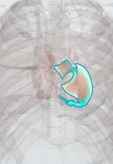
Hypertensive heart disease is the damage that years of high blood pressure do to the heart: a thickened left ventricle that can lead to heart failure.
Updated 2026-10-29
Structures involved
Tap one to see what it is and open it in 3D.
- Left ventricle
- Interventricular septum
- Left atrium
- Ascending aorta
- Aortic arch
- LAD (anterior descending artery)
- Circumflex artery
In biochemistry
In the clinical exam
- Heart sounds: S1, S2 and its splitting, S3 and S4
- Heart failure syndrome: left- and right-sided signs
- Hypertensive emergency: target-organ signs
In physiology
- Heart sounds and where they come from
- Heart failure physiology
- The cardiac electrical axis
- Physiology of hypertension
Why it happens
- Hypertrophy: physiologic and pathologic
- Preload, afterload and contractility
- Cardiac muscle: intercalated discs
What it is
The changes that long-standing high blood pressure causes in the heart: left ventricular hypertrophy, diastolic dysfunction, left atrial enlargement, coronary disease and, eventually, heart failure.
Who gets it
Hypertension is very common in Colombia and Latin America and often poorly controlled, which makes hypertensive heart disease a frequent cause of heart failure and atrial fibrillation.
Causes
- Chronic, poorly controlled arterial hypertension (primary or secondary)
Risk factors
- Longer duration and higher level of hypertension
- Obesity and high salt intake
- Diabetes and chronic kidney disease
- Older age
- Physical inactivity and alcohol
Pathophysiology
The left ventricle faces a constantly high afterload and thickens (concentric hypertrophy) to keep wall stress normal. The thick, fibrotic wall relaxes poorly, so filling pressures rise and the left atrium enlarges. Thicker muscle needs more oxygen while small coronary vessels are damaged, causing ischaemia; over time the ventricle can dilate and lose contraction.
Classification
- Left ventricular hypertrophy without symptoms
- Diastolic dysfunction / heart failure with preserved ejection fraction
- Heart failure with reduced ejection fraction (late, dilated stage)
- Associated atrial fibrillation or coronary ischaemia
Symptoms and signs
- Often none for years
- Breathlessness on exertion, orthopnoea
- Palpitations (atrial fibrillation)
- Chest pain from ischaemia
- Fourth heart sound and a sustained apex beat on examination
Diagnosis
- Clinical
- History of hypertension, repeated blood pressure readings (including home or ambulatory monitoring), and examination for heart failure and target-organ damage.
- Tests
- ECG for left ventricular hypertrophy (voltage criteria such as Sokolow-Lyon), strain pattern or atrial fibrillation. Kidney function, urine albumin, electrolytes, glucose and lipids; natriuretic peptides if heart failure is suspected.
- Imaging
- Echocardiography confirms wall thickening, measures left ventricular mass, diastolic function, left atrial size and ejection fraction. Cardiac MRI helps tell it apart from other causes of hypertrophy.
Differential diagnosis
- Hypertrophic cardiomyopathy
- Aortic stenosis
- Infiltrative disease such as cardiac amyloidosis
- Athlete's heart
- Coronary artery disease
Goal of treatment
Lower blood pressure to target to stop and partly reverse hypertrophy, and prevent heart failure, atrial fibrillation, stroke and coronary events.
Treatment
- Medical
- Antihypertensive drugs, often combined: ACE inhibitors or angiotensin receptor blockers, calcium channel blockers and thiazide-type diuretics; renin–angiotensin blockers help regress hypertrophy.
- Lifestyle
- Less salt, weight loss, regular exercise, limited alcohol and no smoking.
- Medical
- When heart failure or atrial fibrillation appears, they are treated by their own guidelines.
- Follow-up
- Regular blood pressure, kidney and ECG or echo follow-up to check targets and organ damage.
Complications
- Heart failure (preserved, then reduced ejection fraction)
- Atrial fibrillation and stroke
- Myocardial ischaemia and infarction
- Ventricular arrhythmias and sudden death
- Aortic dissection
Prognosis
Left ventricular hypertrophy is a strong independent predictor of cardiovascular events. Good blood pressure control can regress hypertrophy and clearly lowers risk.
Prevention
- Screening and early control of hypertension
- Healthy diet with less salt
- Physical activity and weight control
Test yourself
Think of the answer, then tap the question to see it.
What type of left ventricular hypertrophy does chronic hypertension typically cause?
Concentric hypertrophy: a thicker wall with a normal or small cavity, in response to pressure overload.
Which heart sound suggests a stiff, hypertrophied left ventricle?
A fourth heart sound (S4), from atrial contraction against a non-compliant ventricle.
What is the best simple imaging test to confirm left ventricular hypertrophy?
Echocardiography; the ECG is specific but not very sensitive for hypertrophy.
Which type of heart failure is most typical early in hypertensive heart disease?
Heart failure with preserved ejection fraction, due to diastolic dysfunction.
Which antihypertensive drug classes are especially good at regressing hypertrophy?
Renin–angiotensin system blockers (ACE inhibitors, angiotensin receptor blockers), often with calcium channel blockers.
Related conditions
- Heart failure
- Atrial fibrillation
- Stable angina
- Myocardial Infarction (Heart Attack)
- Arterial hypertension
- Ischaemic stroke
- Intracerebral haemorrhage
- Chagas cardiomyopathy
Educational information, not medical advice. For any symptom, see a health professional.
Sources: Hypertensive Heart Disease (StatPearls (NCBI Bookshelf), 2025).
Spotted a mistake?We are always improving for the community. Tell us what is wrong and our team will review it.Report a mistake
Representative model, for educational purposes only.