Cardiac arrest (VF, pulseless VT, asystole, PEA) !
Cardiac arrest is the sudden stop of effective heart pumping. Without CPR and, when indicated, a defibrillator shock, death follows within minutes.
Updated 2026-11-01
Structures involved
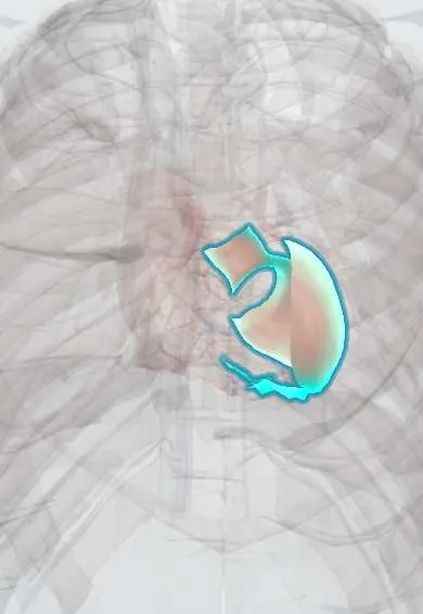
Tap one to see what it is and open it in 3D.
- Left ventricle
- Sinoatrial (SA) node
- LAD (anterior descending artery)
- Right coronary artery
- Right ventricle
- Atrioventricular (AV) node
- Pericardium
Why it happens
- Watershed areas and the organs most sensitive to ischemia
- Hypoxia, ischemia and reperfusion injury
- Mechanical and electrical injury: abrasion, contusion, laceration, wounds
- How arrhythmias arise
- The ventricular action potential
- ECG basics: waves, intervals and leads
What it is
Sudden loss of effective cardiac mechanical activity, with no pulse, no normal breathing and loss of consciousness. The four arrest rhythms are ventricular fibrillation (VF), pulseless ventricular tachycardia (VT), asystole and pulseless electrical activity (PEA).
Who gets it
A leading cause of death worldwide; most out-of-hospital arrests in adults are due to coronary disease. In Colombia and Latin America, survival depends heavily on bystander CPR and public access to automated external defibrillators, which are still limited outside large cities; Chagas cardiomyopathy and rheumatic heart disease add regional causes.
Causes
- Acute coronary ischaemia or scar from a previous myocardial infarction (the most common adult cause)
- Cardiomyopathies (hypertrophic, dilated, Chagas) and inherited channelopathies (long QT, Brugada)
- The reversible «H's and T's»: hypoxia, hypovolaemia, hydrogen ion (acidosis), hypo/hyperkalaemia, hypothermia; tension pneumothorax, tamponade, toxins, thrombosis (pulmonary or coronary)
- Drowning, trauma, airway obstruction and drug overdose, especially in children and young adults
Risk factors
- Known coronary artery disease or previous myocardial infarction
- Reduced left ventricular ejection fraction or heart failure
- Family history of sudden death at a young age
- Electrolyte disorders, QT-prolonging drugs and stimulant use (cocaine)
Pathophysiology
In VF and pulseless VT the ventricles depolarise chaotically or so fast that they do not fill or eject. In PEA there is organised electrical activity without an effective contraction; in asystole there is no electrical activity at all. Brain cells begin to die within minutes without flow, so each minute without CPR or defibrillation sharply lowers survival.
Classification
- Shockable rhythms: ventricular fibrillation and pulseless ventricular tachycardia
- Non-shockable rhythms: asystole and pulseless electrical activity
- Out-of-hospital versus in-hospital cardiac arrest
Symptoms and signs
- Sudden collapse and unresponsiveness
- No pulse (carotid or femoral) and no normal breathing, or only gasping (agonal breathing)
- Sometimes preceded by chest pain, palpitations, dyspnoea, dizziness or fainting
Diagnosis
- Clinical
- Recognised at the bedside: unresponsive patient with no normal breathing and no definite pulse within about 10 seconds. CPR is started at once and the rhythm is checked on the monitor or defibrillator.
- Tests
- Rhythm on the monitor or defibrillator pads; arterial blood gas, potassium, glucose and lactate; troponin and a 12-lead ECG after return of circulation to look for an acute coronary occlusion; end-tidal CO2 to monitor CPR quality.
- Imaging
- Point-of-care ultrasound during pauses can show tamponade, a dilated right ventricle or absent cardiac motion; coronary angiography after return of circulation when an ischaemic cause is likely; brain CT when a neurological cause is suspected.
Differential diagnosis
- Syncope or vasovagal faint with a palpable pulse
- Seizure
- Severe hypoglycaemia
- Opioid or other drug overdose with respiratory arrest but preserved pulse
- Profound shock with a weak pulse that is hard to feel
Goal of treatment
Restore spontaneous circulation as fast as possible with high-quality CPR and early defibrillation when the rhythm is shockable, treat the reversible cause, and protect the brain and heart after return of circulation.
Treatment
- Acute
- Basic life support: call for help, start high-quality chest compressions (adequate rate and depth, full recoil, minimal pauses) with ventilation, and use an automated external defibrillator as soon as it arrives.
- Acute
- Shockable rhythms (VF, pulseless VT): immediate defibrillation, resuming CPR straight after each shock; epinephrine and an antiarrhythmic (amiodarone or lidocaine) when VF persists after shocks.
- Acute
- Non-shockable rhythms (asystole, PEA): CPR with early epinephrine, no shock, and an active search for and treatment of the H's and T's.
- Acute
- Advanced airway when needed, intravenous or intraosseous access, and continuous capnography to guide CPR.
- Follow-up
- Post-arrest care: oxygenation and blood pressure targets, 12-lead ECG and urgent coronary angiography when indicated, temperature control, glucose and seizure monitoring, and a delayed neurological prognosis.
- Procedure
- Implantable cardioverter-defibrillator for survivors of VF or VT arrest without a fully reversible cause.
Complications
- Hypoxic-ischaemic brain injury, the main cause of death after return of circulation
- Post-cardiac arrest syndrome: myocardial dysfunction and systemic inflammatory response
- Rib and sternal fractures from compressions
- Aspiration pneumonia
- Acute kidney injury and multi-organ failure
Prognosis
Survival is low overall and falls with every minute without CPR and defibrillation. Shockable rhythms, a witnessed arrest, bystander CPR and early shock carry the best outcomes; asystole has the worst.
Prevention
- Public CPR training and accessible automated external defibrillators
- Control of coronary risk factors and timely treatment of acute coronary syndromes
- Implantable defibrillator in people at high risk of sudden death
Test yourself
Think of the answer, then tap the question to see it.
Which cardiac arrest rhythms are shockable?
Ventricular fibrillation and pulseless ventricular tachycardia. Asystole and pulseless electrical activity are not shockable.
What are the reversible causes of cardiac arrest summarised as the H's and T's?
Hypoxia, hypovolaemia, hydrogen ion (acidosis), hypo/hyperkalaemia, hypothermia; tension pneumothorax, tamponade, toxins and thrombosis (pulmonary or coronary).
Which two interventions most improve survival from a witnessed VF arrest?
Immediate high-quality chest compressions and early defibrillation.
Which vasopressor is given in both shockable and non-shockable cardiac arrest?
Epinephrine (adrenaline); in non-shockable rhythms it is given as early as possible.
Related conditions
- Ventricular tachycardia
- Myocardial Infarction (Heart Attack)
- Hypertrophic cardiomyopathy
- Dilated cardiomyopathy
- Cardiac tamponade
- Pulmonary embolism
- Pneumothorax
- Hypovolaemic shock
- Atrioventricular (heart) block
Educational information, not medical advice. For any symptom, see a health professional.
Sources: Cardiac arrest: MedlinePlus Medical Encyclopedia (MedlinePlus, US National Library of Medicine, 2026).
Spotted a mistake?We are always improving for the community. Tell us what is wrong and our team will review it.Report a mistake
Representative model, for educational purposes only.