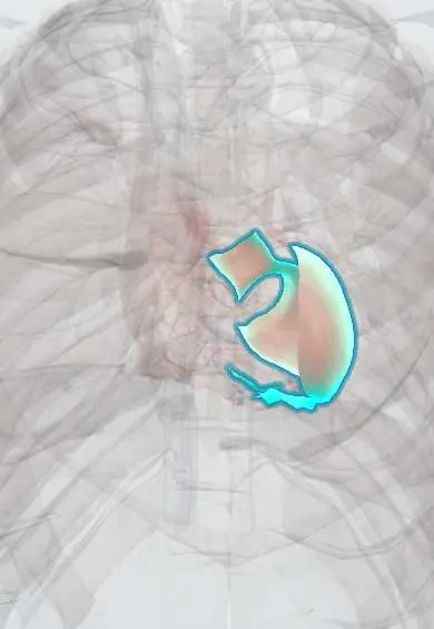
Ventricular tachycardia !
Ventricular tachycardia is a fast rhythm that starts in the lower chambers of the heart. It can cause fainting, cardiac arrest and sudden death.
Updated 2026-11-01
Structures involved
Tap one to see what it is and open it in 3D.
- Left ventricle
- Right ventricle
- Bundle branches
- Interventricular septum
- LAD (anterior descending artery)
- Bundle of His
Why it happens
- The myocardial infarct over time: what the pathologist sees from hours to months
- How arrhythmias arise
- The cardiac conduction system
- The ventricular action potential
- Cardiac muscle: intercalated discs
What it is
A tachycardia of three or more consecutive beats arising below the bundle of His, usually above 100 beats per minute, with wide QRS complexes on the ECG. It may be stable, unstable or pulseless.
Who gets it
Most cases occur in people with structural heart disease, especially scar from a previous infarction. In Colombia and Latin America, chronic Chagas cardiomyopathy is an important and characteristic cause of sustained VT and sudden death.
Causes
- Scar from a previous myocardial infarction (re-entry around the scar), the most common cause of monomorphic VT
- Acute ischaemia, which typically causes polymorphic VT
- Cardiomyopathies: Chagas, dilated, hypertrophic, arrhythmogenic right ventricular
- Long QT (congenital or drug-induced) causing torsades de pointes; hypokalaemia and hypomagnesaemia
- Idiopathic VT in structurally normal hearts (outflow tract, fascicular)
Risk factors
- Previous myocardial infarction and low ejection fraction
- Heart failure
- Electrolyte disorders and QT-prolonging drugs
- Family history of sudden death or inherited channelopathy
Pathophysiology
Most sustained VT is re-entry: an electrical circuit loops through slow-conducting tissue at the edge of a scar. Activation spreads from muscle cell to muscle cell instead of through the His-Purkinje system, so the QRS is wide. The fast rate shortens filling and lowers output, and VT can degenerate into ventricular fibrillation.
Classification
- Non-sustained (under 30 seconds, no compromise) or sustained (30 seconds or more, or needing termination)
- Monomorphic or polymorphic; torsades de pointes is polymorphic VT with a long QT
- Stable with a pulse, unstable with a pulse, or pulseless
Symptoms and signs
- Palpitations
- Dizziness, presyncope or syncope
- Chest pain and dyspnoea
- Hypotension and signs of shock
- Sudden collapse when there is no pulse
Diagnosis
- Clinical
- Check pulse, blood pressure and consciousness first, since stability decides treatment. A wide-complex tachycardia in someone with structural heart disease is treated as VT until proven otherwise; cannon a waves and a variable first heart sound suggest AV dissociation.
- Tests
- 12-lead ECG: wide regular QRS, AV dissociation, capture and fusion beats, concordance in the precordial leads. Potassium, magnesium, troponin; QT interval after conversion; Holter monitoring; Chagas serology in people from endemic areas; electrophysiological study in selected cases.
- Imaging
- Echocardiography to assess ejection fraction and structural disease; cardiac MRI to detect scar or infiltration; coronary angiography when ischaemia is suspected.
Differential diagnosis
- Supraventricular tachycardia with aberrant conduction (bundle branch block)
- Pre-excited tachycardia (Wolff-Parkinson-White)
- Sinus tachycardia with a pre-existing bundle branch block
- Hyperkalaemia or sodium-channel-blocker toxicity with wide QRS
- Pacemaker-mediated tachycardia
Goal of treatment
Terminate the arrhythmia safely according to haemodynamic stability, correct triggers such as ischaemia and electrolytes, and prevent recurrence and sudden death.
Treatment
- Acute
- Pulseless VT: managed as cardiac arrest with CPR and immediate defibrillation.
- Acute
- Unstable VT with a pulse (hypotension, shock, altered mental status, ischaemic pain, heart failure): urgent synchronised cardioversion with sedation when possible.
- Medical
- Stable monomorphic VT: intravenous antiarrhythmics (amiodarone, procainamide or lidocaine) or elective synchronised cardioversion; correction of potassium and magnesium.
- Medical
- Torsades de pointes: intravenous magnesium sulfate, stopping QT-prolonging drugs, and increasing heart rate (pacing or isoproterenol) when it recurs.
- Procedure
- Prevention of sudden death: implantable cardioverter-defibrillator in selected patients; catheter ablation for recurrent VT; long-term beta-blockers or amiodarone.
Complications
- Degeneration into ventricular fibrillation and sudden cardiac death
- Cardiogenic shock
- Syncope with injury
- Worsening heart failure
- Tachycardia-induced cardiomyopathy when incessant
Prognosis
Depends on the underlying heart. Idiopathic VT in a normal heart has a good outlook and often responds to ablation; VT with scar or low ejection fraction carries a high risk of sudden death, which an implantable defibrillator lowers.
Prevention
- Early reperfusion of infarction and optimal heart failure treatment
- Avoiding QT-prolonging drug combinations and correcting electrolytes
- Chagas control and screening in endemic areas
Test yourself
Think of the answer, then tap the question to see it.
How is a wide-complex tachycardia in a patient with a previous infarction managed until proven otherwise?
As ventricular tachycardia.
What is the treatment of unstable VT with a pulse?
Synchronised electrical cardioversion.
Which ECG findings strongly favour VT over SVT with aberrancy?
AV dissociation, capture beats and fusion beats, and precordial concordance.
What is the first-line drug for torsades de pointes?
Intravenous magnesium sulfate, along with stopping QT-prolonging drugs and correcting potassium.
Which parasitic heart disease is a classic cause of VT in Latin America?
Chronic Chagas cardiomyopathy, caused by Trypanosoma cruzi.
Related conditions
- Cardiac arrest (VF, pulseless VT, asystole, PEA)
- Myocardial Infarction (Heart Attack)
- Chagas cardiomyopathy
- Dilated cardiomyopathy
- Hypertrophic cardiomyopathy
- Heart failure
- Supraventricular tachycardia (AVNRT, AVRT)
Educational information, not medical advice. For any symptom, see a health professional.
Sources: Ventricular tachycardia: MedlinePlus Medical Encyclopedia (MedlinePlus, US National Library of Medicine, 2026); Chagas disease (also known as American trypanosomiasis) (World Health Organization, 2026).
Spotted a mistake?We are always improving for the community. Tell us what is wrong and our team will review it.Report a mistake
Representative model, for educational purposes only.