
Intermediate uveitis and pars planitis
Inflammation centred in the vitreous gel causes floaters and blurred vision. It can be linked to multiple sclerosis or sarcoidosis.
What it is
Uveitis centred in the vitreous and peripheral retina, often bilateral and chronic, mostly in children and young adults. With snowbanks or snowballs and no infection or systemic disease, it is called pars planitis. It is linked to multiple sclerosis and sarcoidosis; cystoid macular oedema causes most visual loss.
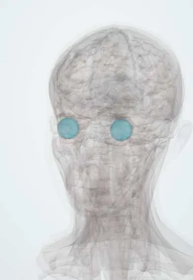
See it in 3D
Who gets it
Accounts for roughly a tenth to a fifth of uveitis; peaks in children and young adults; a few percent to a fifth of patients have or develop multiple sclerosis.
Causes
- Idiopathic (pars planitis) in most cases
- Multiple sclerosis
- Sarcoidosis
- Infections: syphilis, tuberculosis, Lyme disease, toxocariasis
- Intraocular lymphoma in older adults (masquerade)
Risk factors
- Age under 40
- HLA-DR15 (shared with multiple sclerosis)
Pathophysiology
T-cell inflammation of the peripheral retina and ciliary body releases cells into the vitreous. Peripheral venules become sheathed (periphlebitis), inflammatory exudate gathers on the inferior pars plana, and leaky vessels lead to macular oedema.
Symptoms and signs
- Floaters
- Blurred vision
- Little pain or redness
Diagnosis
- Clinical
- Vitreous cells and haze, snowballs (inflammatory clumps) in the inferior vitreous, snowbanks on the inferior pars plana on indirect ophthalmoscopy with indentation, peripheral periphlebitis; mild anterior chamber reaction.
- Tests
- Syphilis serology, interferon-gamma release assay, angiotensin-converting enzyme and lysozyme, Lyme serology where endemic; neurological history and examination before starting anti-TNF therapy.
- Imaging
- OCT for macular oedema; fluorescein angiography for leakage and peripheral vasculitis; chest imaging for sarcoidosis; brain MRI when neurological symptoms suggest multiple sclerosis.
Differential diagnosis
- Sarcoid uveitis
- Multiple sclerosis-associated uveitis
- Intraocular lymphoma
- Toxocariasis
- Fuchs uveitis syndrome
Vitreous cells in a white eye: what else it could be
| Feature | Intermediate uveitis / pars planitis | Vitreoretinal lymphoma | Fuchs uveitis | Sarcoid uveitis |
|---|---|---|---|---|
| Usual age | Children and young adults | Usually over 50 | 20 to 50 | Adults of any age |
| Laterality | Often both eyes | Often both eyes | Usually one eye | Often both eyes |
| Key signs | Inferior snowballs and snowbanks, periphlebitis | Sheets of vitreous cells, creamy subretinal infiltrates | Stellate keratic precipitates over the whole cornea, iris atrophy | Granulomatous keratic precipitates, iris nodules, periphlebitis, snowballs |
| Steroid response | Responds | Partial or short-lived | Little effect | Responds |
| Key test | OCT of the macula; brain MRI if neurological symptoms | Vitreous biopsy and brain MRI | Clinical; aqueous rubella antibodies if doubtful | Chest imaging, angiotensin-converting enzyme, lysozyme |
Goal of treatment
Treat when vision is threatened, mainly to clear macular oedema, and screen for associated disease.
Treatment
- Follow-up
- Mild disease with good vision and no macular oedema can be observed.
- Medical
- Periocular or intravitreal corticosteroids, then systemic corticosteroids and steroid-sparing agents (methotrexate, mycophenolate, adalimumab). Anti-TNF drugs are avoided in multiple sclerosis because they can worsen demyelination; interferon beta can be used instead.
- Procedure
- Peripheral laser photocoagulation or cryotherapy of the snowbank in selected cases.
- Surgical
- Pars plana vitrectomy for persistent opacities, vitreous haemorrhage or traction.
Complications
- Cystoid macular oedema
- Cataract
- Glaucoma
- Epiretinal membrane
- Vitreous haemorrhage and retinal detachment
- Peripheral neovascularisation
Common traps
- Floaters in a young person with a white eye are harmless vitreous change: vitreous cells under 40 suggest intermediate uveitis, which needs a dilated exam with indentation to find snowbanks
- Pars planitis is just another name for any intermediate uveitis: the label is used only when snowbanks or snowballs are present and no infection or systemic disease is found
- Anti-TNF is a safe step-up for every patient: it can trigger or worsen demyelination, so ask about neurological symptoms and image the brain when they suggest multiple sclerosis
- Visual loss here comes from the vitreous haze: most lasting loss comes from cystoid macular oedema, so the macula is checked with OCT at every visit
- New intermediate uveitis after 50 is late pars planitis: think of vitreoretinal lymphoma masquerading as uveitis, especially if steroids help only briefly
Prognosis
Long course with flares, but most patients keep good vision when macular oedema is treated.
Test yourself
Think of the answer, then tap the question to see it.
A 12-year-old has floaters in both white eyes; indentation shows grey-white exudate on the inferior pars plana, and work-up is negative. Which diagnosis?
Pars planitis, the idiopathic form of intermediate uveitis with snowbanks.
Why do snowballs and snowbanks collect in the lower part of the eye?
Inflammatory cells and exudate in the vitreous settle with gravity onto the inferior vitreous base and pars plana.
A 25-year-old with intermediate uveitis needs a steroid-sparing biologic and had arm numbness last year. What comes before an anti-TNF?
A neurological history and examination and brain MRI, because anti-TNF can worsen demyelination; if multiple sclerosis is present, an option such as interferon beta is chosen.
When is mild intermediate uveitis treated rather than observed?
When vision is threatened, mainly by cystoid macular oedema, or by complications such as dense haze, vitreous haemorrhage or neovascularisation.
A 68-year-old develops new vitreous cells that improve only briefly with steroids. What must be excluded, and how?
Vitreoretinal lymphoma masquerading as uveitis; a vitreous biopsy and brain MRI are needed.
Connections
Educational information, not medical advice. For any symptom, see a health professional.
Sources: Pars Planitis (StatPearls Publishing (NCBI Bookshelf), 2023); Intermediate Uveitis (EyeWiki (American Academy of Ophthalmology), 2025).
Spotted a mistake?We are always improving for the community. Tell us what is wrong and our team will review it.Report a mistake
Representative model, for educational purposes only.