
Vitreoretinal lymphoma
A lymphoma inside the eye that looks like stubborn uveitis in an older adult. Many patients also have or develop lymphoma in the brain.
What it is
An aggressive non-Hodgkin lymphoma, usually diffuse large B-cell, in the vitreous and under the retina. It is the classic 'masquerade syndrome': an older adult's 'uveitis' that responds poorly or briefly to corticosteroids. A subtype of primary CNS lymphoma, it involves the brain in most patients.
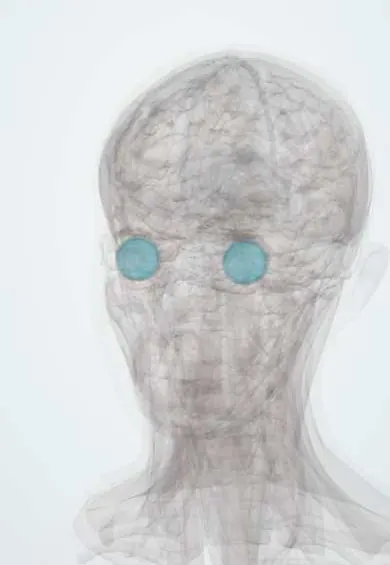
See it in 3D
Who gets it
Rare; usually over 50 years, with a slight female predominance. Bilateral in most patients eventually. Brain involvement occurs in a majority over time, and a smaller proportion of brain lymphomas involve the eye.
Causes
- Malignant B lymphocytes (diffuse large B-cell lymphoma), often with MYD88 L265P mutation
- Rarely T-cell lymphoma or spread from systemic lymphoma
Risk factors
- Age over 50
- Immunosuppression (HIV, organ transplant)
- Previous primary central nervous system lymphoma
Pathophysiology
Lymphoma cells home to the vitreous and to the space between the retinal pigment epithelium and Bruch membrane. They cause sheets of vitreous cells with little true inflammation, and creamy-yellow sub-RPE infiltrates. Corticosteroids kill some tumour cells, which is why the 'uveitis' briefly improves and why biopsy should be taken before steroids.
Symptoms and signs
- Floaters
- Painless blurred vision
- Usually a white, quiet eye
- Neurological symptoms (personality change, headache, weakness) if the brain is involved
Diagnosis
- Clinical
- Slit-lamp and fundus examination: dense vitreous cells in sheets or 'aurora borealis' strands, little anterior inflammation, sub-RPE infiltrates with 'leopard spot' pigment.
- Tests
- Diagnostic vitrectomy (ideally before corticosteroids) for cytology, flow cytometry, IL-10 to IL-6 ratio over 1 and MYD88 testing; cerebrospinal fluid cytology; HIV test.
- Imaging
- OCT shows hyper-reflective deposits under the RPE; fluorescein angiography and autofluorescence show granular 'leopard spot' changes; MRI of the brain with contrast for CNS disease.
Differential diagnosis
- Chronic intermediate or posterior uveitis
- Sarcoidosis
- Syphilis and tuberculosis
- Acute retinal necrosis
- Birdshot chorioretinopathy
- Endophthalmitis
Vitreous cells in an adult: inflammation or lymphoma?
| Condition | Patient | Eye findings | Response to corticosteroids | Key test |
|---|---|---|---|---|
| Vitreoretinal lymphoma | Over 50 | White eye, sheets of vitreous cells, creamy sub-RPE infiltrates | Brief or partial, then relapse | Diagnostic vitrectomy (cytology, flow cytometry, IL-10 to IL-6 ratio, MYD88) |
| Intermediate uveitis | Child or young adult | Snowballs, pars plana snowbank, macular oedema | Good and lasting | Exclude sarcoidosis, multiple sclerosis, tuberculosis, syphilis |
| Ocular sarcoidosis | Any adult | Granulomatous keratic precipitates, iris nodules, candle-wax periphlebitis | Good | Chest imaging, ACE and lysozyme |
| Acute retinal necrosis | Any age | Painful red eye, spreading peripheral white retinal necrosis | Worsens without antivirals | PCR of aqueous or vitreous for herpesviruses |
Goal of treatment
Control the eye disease to save vision and treat or prevent brain lymphoma with oncology.
Treatment
- Medical
- Intravitreal methotrexate or rituximab for eye disease; systemic high-dose methotrexate-based chemotherapy with oncology when the brain is involved.
- Procedure
- Ocular external beam radiotherapy as an alternative or for bilateral disease.
- Follow-up
- Close follow-up with repeated brain MRI because brain lymphoma often appears later.
Complications
- Central nervous system lymphoma
- Retinal detachment
- Optic nerve infiltration
- Loss of vision
- Treatment toxicity (corneal epitheliopathy from intravitreal drugs, radiation retinopathy)
Common traps
- Vitreous cells always mean uveitis: in an adult over 50 with a white, quiet eye and steroid-resistant vitritis, lymphoma is the masquerade to exclude
- A good response to corticosteroids rules out lymphoma: steroids kill lymphoma cells, so the eye improves briefly and a later biopsy may be falsely negative
- Once the eye is controlled the patient is cured: most patients develop brain lymphoma, so brain MRI and cerebrospinal fluid are checked during follow-up
- A negative vitreous cytology excludes the diagnosis: lymphoma cells are few and fragile, so flow cytometry, the IL-10 to IL-6 ratio and MYD88 testing add sensitivity, and repeat sampling may be needed
Prognosis
Eye disease responds well, but overall survival is limited by brain involvement, with median survival of a few years.
Test yourself
Think of the answer, then tap the question to see it.
A 68-year-old's 'uveitis' relapses each time corticosteroids are tapered; the eye stays white. What next?
Suspect vitreoretinal lymphoma: hold further steroids, arrange diagnostic vitrectomy, contrast brain MRI and cerebrospinal fluid cytology.
Why does the 'uveitis' briefly improve with corticosteroids?
Corticosteroids kill some lymphoma cells, so the vitreous clears for a while before the tumour returns.
Why is brain MRI repeated even after the eye disease is controlled?
It is a subtype of primary CNS lymphoma, and most patients develop brain disease over time.
Which vitreous cytokine pattern points to lymphoma rather than inflammation?
IL-10 higher than IL-6 (ratio over 1): lymphoma B cells secrete IL-10, while inflammation mainly raises IL-6.
Why it happens
In haematology
Connections
Educational information, not medical advice. For any symptom, see a health professional.
Sources: Primary Intraocular Lymphoma (StatPearls Publishing (NCBI Bookshelf), 2026); Primary Vitreoretinal Lymphoma (EyeWiki (American Academy of Ophthalmology), 2025).
Spotted a mistake?We are always improving for the community. Tell us what is wrong and our team will review it.Report a mistake
Representative model, for educational purposes only.