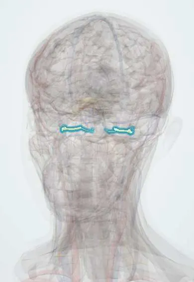
Nervous system · Head and neck
Vestibulocochlear nerve (VIII) !
The vestibulocochlear nerve (cranial nerve VIII) carries hearing and balance from the inner ear to the brainstem.
Official name: Vestibulocochlear nerve
Opens in the Durgical app, no sign-up needed.
What it is
The nerve from the inner ear to the brainstem, with hearing and balance parts.
What it does
Hearing and balance; a benign tumour on it (acoustic neuroma) causes one-sided hearing loss.
Key facts
- Roots
- Cochlear and vestibular nuclei (pons/medulla); internal acoustic meatus; sensory ganglia: spiral (in the modiolus) and vestibular (Scarpa, in the meatus)
- Moves
- None
- Feeling from
- Hearing (cochlear part); balance (vestibular part)
Relations
- Course
- From the cochlea and the vestibular sense organs through the internal acoustic meatus, then across the cerebellopontine angle cistern to the pontomedullary junction
- In front
- Facial nerve and nervus intermedius, which travel with it through the internal acoustic meatus
- Below
- Loops of the anterior inferior cerebellar artery in the angle, giving the labyrinthine artery that runs with the nerve
- Behind
- Flocculus of the cerebellum and the lateral recess of the fourth ventricle
How to find it
Seen on MRI or at surgery in the cerebellopontine angle, between the pons and the internal acoustic meatus, below the trigeminal nerve and above the glossopharyngeal and vagus nerves.
Examination
- Rinne and Weber: a sensorineural pattern on the affected side (Rinne positive, Weber to the other ear)
- Audiometry: speech discrimination worse than the pure-tone loss suggests a retrocochlear (nerve) lesion
- Auditory brainstem responses: delayed waves on one side point to a lesion of the nerve
- Head impulse and caloric tests examine the vestibular part
On imaging
Gadolinium-enhanced T1 MRI or high-resolution heavily T2-weighted MRI of the internal acoustic meatus: a schwannoma enhances and fills the meatus, widening into the angle in an «ice cream cone» shape.
Common problems
- Vestibular schwannoma (acoustic neuroma): a slow benign tumour causing one-sided hearing loss, ringing and imbalance.
How it is diagnosed
Hearing test and MRI with contrast.
Treatment
- Watching small tumours.
- Radiosurgery or microsurgery for growing ones.
Clinical notes
- Large schwannomas press on the trigeminal nerve (loss of the corneal reflex is an early sign), the facial nerve, the cerebellum and the fourth ventricle (hydrocephalus)
- Surgery or radiosurgery for schwannoma puts the facial nerve at risk because it runs on the tumour surface
- Vestibular neuritis is thought to be viral inflammation, usually of the superior vestibular nerve
Anatomical variants
- A loop of the anterior inferior cerebellar artery often reaches the opening of the internal acoustic meatus or enters it; on MRI it is usually a harmless finding
Development
The inner ear and the neurons of the cochlear and vestibular ganglia come from the otic placode, a thickening of surface ectoderm beside the hindbrain that sinks in to form the otic vesicle.
Memory aid
In the internal acoustic meatus the facial nerve (VII) runs on top and the cochlear nerve below it, both in front; the two vestibular nerves lie behind them.
Related conditions
- Vestibular schwannoma (acoustic neuroma)
- Presbycusis (age-related hearing loss)
- Neurofibromatosis (types 1 and 2)
- Hearing loss (conductive and sensorineural, including sudden hearing loss)
- Vestibular neuritis and labyrinthitis
Test yourself
Think of the answer, then tap the question to see it.
From which cells does a vestibular schwannoma (acoustic neuroma) arise, and what do bilateral tumours define?
Schwann cells of the vestibular nerve in the internal acoustic meatus; bilateral tumours define neurofibromatosis type 2.
How are the nerves arranged in the internal acoustic meatus?
Facial nerve anterosuperior, cochlear nerve anteroinferior, superior and inferior vestibular nerves posterior.
What is an early sign of a large vestibular schwannoma pressing on the trigeminal nerve?
Loss of the corneal reflex on that side.
What do the Rinne and Weber tests show with a vestibulocochlear nerve lesion?
A sensorineural pattern: Rinne positive on the affected side and Weber lateralising to the other ear.
Educational information, not medical advice. For any symptom, see a health professional.
Its names
- Official Latin (TA2)
- nervus vestibulocochlearis TA2 6307
- Official Spanish (DeCS)
- Nervio Vestibulococlear DeCS D000159
- English (TA2)
- Vestibulocochlear nerve
- Also called
- acoustic nerve · cranial nerve VIII · nervus acusticus · nervus statoacusticus · nervus octavus
Related
Sources: Terminologia Anatomica (TA2); DeCS; Neuroanatomy, Cranial Nerve 8 (Vestibulocochlear).
Spotted a mistake?We are always improving for the community. Tell us what is wrong and our team will review it.Report a mistake
Representative model, for educational purposes only.