Conditions · Pediatrics and neonatology
Necrotising enterocolitis !
A dangerous bowel disease of premature babies in which part of the intestine becomes inflamed and dies. It needs urgent care.
Updated 2026-11-01
Structures involved
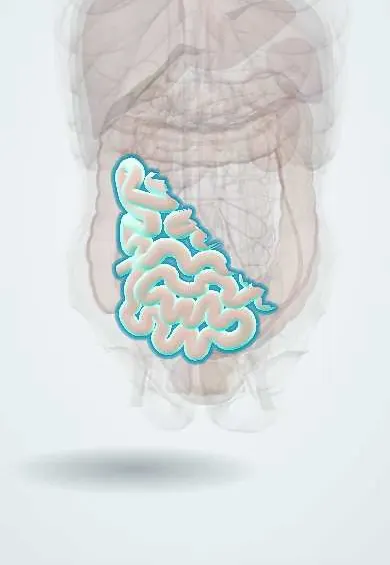
Tap one to see what it is and open it in 3D.
In embryology
Why it happens
- Patterns of necrosis: coagulative, liquefactive, caseous, fat, fibrinoid, gangrenous
- Necrotizing enterocolitis: why the premature gut is vulnerable
- Prematurity and fetal growth restriction
- Splanchnic and portal circulation
- Small intestine: villi, crypts, enterocytes, Paneth cells; duodenum vs jejunum vs ileum
What it is
An acute inflammatory and ischaemic necrosis of the intestinal wall, typically of the terminal ileum and proximal colon, that mainly affects preterm infants after feeds have started. It is the most common gastrointestinal emergency of the neonatal period.
Who gets it
It affects a few to about 10% of very-low-birth-weight infants and is much rarer at term. It usually appears in the second to third week of life in very preterm babies. In Latin American neonatal units it is a frequent cause of surgery and death among preterm infants.
Causes
- Multifactorial: intestinal immaturity, abnormal bacterial colonisation and enteral feeding acting together
- In term infants, usually linked to reduced gut blood flow (congenital heart disease, polycythaemia, asphyxia)
Risk factors
- Prematurity and very low birth weight (the main risk factor)
- Formula feeding instead of human milk
- Rapid advancement of feeds
- Perinatal asphyxia, hypotension or congenital heart disease
- Prolonged empirical antibiotics and acid-suppressing drugs
- Red cell transfusion and severe anaemia (proposed association)
Pathophysiology
An immature intestinal barrier, poor motility and an exaggerated inflammatory response in the preterm gut meet dysbiotic bacteria and substrate from feeds. Bacterial translocation and inflammation cause mucosal injury and ischaemia that can extend through the wall; gas produced by bacteria dissects into the wall (pneumatosis intestinalis) and the portal venous system, and transmural necrosis leads to perforation and peritonitis.
Classification
- Modified Bell stage I (suspected): non-specific systemic and mild abdominal signs
- Bell stage II (definite): pneumatosis intestinalis on X-ray, with or without portal venous gas
- Bell stage III (advanced): severe illness with shock and peritonitis, with intact bowel (IIIA) or perforation (IIIB)
Symptoms and signs
- Feeding intolerance with increased gastric residuals or vomiting, often bilious
- Abdominal distension and tenderness
- Blood in the stools (visible or occult)
- Lethargy, apnoea, bradycardia and temperature instability
- Abdominal wall erythema or discolouration in advanced disease
Diagnosis
- Clinical
- A preterm infant on enteral feeds with feeding intolerance, abdominal distension and bloody stools; staging with modified Bell criteria guides treatment, with serial abdominal examinations.
- Tests
- Blood count (low or high white cells, falling platelets), C-reactive protein, blood gases (metabolic acidosis), electrolytes, coagulation and blood cultures.
- Imaging
- Abdominal X-ray (supine and left lateral decubitus or cross-table) is key: dilated loops, pneumatosis intestinalis (pathognomonic), portal venous gas and pneumoperitoneum when perforated; repeated every few hours in the acute phase. Abdominal ultrasound shows bowel wall perfusion, thickness and free fluid.
Differential diagnosis
- Neonatal sepsis with ileus
- Spontaneous intestinal perforation (usually extremely preterm, without pneumatosis)
- Malrotation with midgut volvulus
- Hirschsprung enterocolitis
- Cow's milk protein-induced enterocolitis
Goal of treatment
Rest the bowel, decompress it and treat infection and shock early to stop progression, and remove necrotic bowel or drain the abdomen when perforation or failure of medical treatment occurs.
Treatment
- Medical
- Stop enteral feeds, decompress the stomach with an orogastric tube, give parenteral nutrition and broad-spectrum intravenous antibiotics (including anaerobic cover in advanced disease) for about one to two weeks depending on stage.
- Acute
- Supportive care for shock, acidosis, coagulopathy and respiratory failure: fluids, vasoactive drugs, blood products and ventilation.
- Surgical
- Laparotomy with resection of necrotic bowel and stoma, or bedside peritoneal drainage in very small unstable infants, when there is perforation or deterioration despite medical treatment.
- Follow-up
- Nursing care: measure abdominal girth, check gastric residuals and stools for blood, monitor vital signs and perfusion, keep the infant supine without nappy pressure on the abdomen, and reintroduce feeds slowly with human milk.
Complications
- Intestinal perforation, peritonitis and septic shock
- Intestinal strictures, mainly of the colon, causing later obstruction
- Short bowel syndrome and intestinal failure after extensive resection
- Cholestasis linked to long parenteral nutrition
- Neurodevelopmental impairment and death
Prognosis
Mortality is substantial and highest in the smallest infants and those needing surgery; survivors may have strictures, short bowel or delayed development, while medically treated mild cases usually recover fully.
Prevention
- Exclusive human milk, the mother's own or donor milk
- Standardised feeding protocols with careful advancement
- Antenatal corticosteroids and prevention of preterm birth
- Limiting unnecessary antibiotics and acid suppressants; probiotics in some units
Test yourself
Think of the answer, then tap the question to see it.
What is the main risk factor for necrotising enterocolitis?
Prematurity, especially very low birth weight.
What is the pathognomonic X-ray sign of NEC?
Pneumatosis intestinalis: gas within the bowel wall.
Which part of the bowel is most often affected in NEC?
The terminal ileum and proximal (ascending) colon.
What is the initial medical management of NEC?
Stop feeds, orogastric decompression, parenteral nutrition and broad-spectrum intravenous antibiotics, with serial X-rays.
What is the main absolute indication for surgery in NEC?
Intestinal perforation (pneumoperitoneum).
Related conditions
- Prematurity and its complications
- Neonatal sepsis
- Peritonitis
- Small bowel obstruction
- Intussusception (children)
Educational information, not medical advice. For any symptom, see a health professional.
Sources: Necrotizing enterocolitis (MedlinePlus (U.S. National Library of Medicine), 2024).
Spotted a mistake?We are always improving for the community. Tell us what is wrong and our team will review it.Report a mistake
Representative model, for educational purposes only.