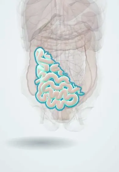
Intussusception (children) !
Intussusception is when one part of the bowel slides into the next like a telescope; it is the commonest cause of bowel blockage in babies and toddlers.
Updated 2026-10-29
Structures involved
Tap one to see what it is and open it in 3D.
- Ileum
- Cecum
- Ascending colon
- Appendix
- Mesenteric lymph nodes
- Ileocolic artery
- Mesenteric veins
- Transverse colon
In immunology
In embryology
In physiology
Why it happens
What it is
Telescoping of one segment of bowel into the next, most often the end of the ileum into the colon (ileocolic). It is the commonest cause of bowel obstruction in infants and toddlers and a surgical emergency.
Who gets it
Most cases occur between about 6 months and 3 years of age, peaking in infancy, and boys are affected more often. Cases follow viral infections. In adults it is rare and usually caused by a tumour.
Causes
- Idiopathic in most children: swollen Peyer patches (lymphoid hyperplasia) after a viral infection act as the lead point
- Pathological lead points, more likely outside the usual age: Meckel diverticulum, polyps, lymphoma, duplication cysts
- IgA vasculitis (Henoch–Schönlein purpura): bowel wall haematoma, often small bowel into small bowel
- Cystic fibrosis
Risk factors
- Age 6 months to 3 years
- Male sex
- Recent viral gastroenteritis or respiratory infection
- A small rise in risk after rotavirus vaccination, far outweighed by its benefits
- Previous intussusception
Pathophysiology
Peristalsis drags the proximal bowel (intussusceptum) with its mesentery into the distal bowel (intussuscipiens). The mesentery is compressed, so venous and lymphatic return are blocked first: the bowel swells and the mucosa bleeds, producing mucus mixed with blood. Without reduction the arterial supply fails, leading to ischaemia, necrosis and perforation.
Classification
- Ileocolic (most common), ileoileal or colocolic
- Idiopathic or with a pathological lead point
- Short transient small-bowel intussusception seen on ultrasound, which often resolves by itself
Symptoms and signs
- Sudden bouts of colicky abdominal pain: the child screams and draws up the legs, then settles between attacks
- Vomiting, becoming bilious as obstruction progresses
- Red-currant jelly stool (blood and mucus), often a late sign
- Sausage-shaped mass in the right or upper abdomen, with an empty right lower quadrant
- Lethargy or drowsiness, sometimes the only sign in infants
Diagnosis
- Clinical
- Suspected in an infant or toddler with intermittent inconsolable crying, vomiting and lethargy; the classic triad of colicky pain, a mass and red-currant jelly stool is present in only a minority.
- Tests
- Blood tests are nonspecific: electrolytes and blood gas for dehydration and acidosis, blood count, and blood group before reduction or surgery.
- Imaging
- Ultrasound is the test of choice: a target (doughnut) sign in transverse view and a pseudokidney sign in longitudinal view. A plain abdominal X-ray may show obstruction or free air but cannot rule it out.
Differential diagnosis
- Acute gastroenteritis
- Infant colic
- Malrotation with midgut volvulus
- Incarcerated inguinal hernia
- Meckel diverticulum
- Appendicitis or IgA vasculitis
Goal of treatment
Rapid fluid resuscitation and reduction of the intussusception before the bowel becomes ischaemic, with surgery when reduction fails or the child has peritonitis.
Treatment
- Acute
- Intravenous fluid resuscitation, nasogastric decompression if vomiting, analgesia and early surgical assessment.
- Procedure
- Image-guided enema reduction with air (pneumatic) or with saline or contrast (hydrostatic), under fluoroscopy or ultrasound, in stable children without peritonitis; it succeeds in most cases.
- Surgical
- Surgery (laparoscopic or open) after a failed enema or with perforation, peritonitis or shock: manual reduction, or resection of non-viable bowel or a lead point.
- Follow-up
- Observation after reduction, because recurrence can occur, usually soon after.
Complications
- Bowel ischaemia and necrosis
- Perforation during the illness or, rarely, during the enema
- Peritonitis and sepsis
- Hypovolaemic shock
- Recurrence
Prognosis
Excellent with early diagnosis and reduction; delay increases the risk of bowel resection and death.
Test yourself
Think of the answer, then tap the question to see it.
What is the most common type of intussusception in children?
Ileocolic: the end of the ileum telescopes into the cecum and colon.
What is the classic ultrasound sign of intussusception?
The target (doughnut) sign in transverse view; the pseudokidney sign in longitudinal view.
What is red-currant jelly stool?
Stool made of blood and mucus from congested, ischaemic bowel mucosa; it is a late sign.
What is the first-line treatment for a stable child without peritonitis?
Image-guided air (pneumatic) or hydrostatic enema reduction.
What is the usual lead point in idiopathic childhood intussusception?
Enlarged Peyer patches (lymphoid tissue) after a viral infection.
Related conditions
- Small bowel obstruction
- Acute appendicitis
- Inguinal hernia
- Acute (viral) gastroenteritis
- Rectal prolapse
Educational information, not medical advice. For any symptom, see a health professional.
Sources: Child Intussusception (StatPearls (NCBI Bookshelf), 2023).
Spotted a mistake?We are always improving for the community. Tell us what is wrong and our team will review it.Report a mistake
Representative model, for educational purposes only.