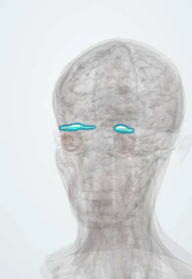
Myasthenia gravis !
Myasthenia gravis is an autoimmune disease in which antibodies block nerve-to-muscle signals, causing weakness that worsens with use, often droopy eyelids.
Updated 2026-11-01
Structures involved
Tap one to see what it is and open it in 3D.
- Levator palpebrae
- Lateral rectus
- Thymus
- Medial rectus
- Superior rectus
- Inferior rectus
- Orbicularis oculi
- Masseter
- Sternocleidomastoid
- Diaphragm
In histology
In the clinical exam
- Eye exam: visual acuity, visual fields and eye movements
- Cranial nerve examination
- Motor examination: bulk, tone and strength grading
Why it happens
What it is
A chronic autoimmune disease of the neuromuscular junction in which antibodies against the acetylcholine receptor (or related proteins) cause fluctuating weakness of voluntary muscles that gets worse with use and improves with rest. Eye, face, swallowing and breathing muscles are often involved.
Who gets it
The most common disorder of neuromuscular transmission. It has two peaks: women in their 20s and 30s and men over 60. It occurs worldwide, including Colombia and Latin America; about 10–15% of patients have a thymoma and many others have thymic hyperplasia.
Causes
- Antibodies against the acetylcholine receptor in most generalised cases
- Antibodies against muscle-specific kinase (MuSK) or LRP4 in some seronegative cases
- Thymic abnormalities: hyperplasia or thymoma
- Rarely triggered by drugs such as immune checkpoint inhibitors or penicillamine
Risk factors
- Female sex in younger adults, male sex in older adults
- Other autoimmune diseases (autoimmune thyroid disease, lupus, rheumatoid arthritis)
- Thymoma
Pathophysiology
IgG antibodies bind postsynaptic acetylcholine receptors, block them, speed their removal and activate complement that damages the muscle membrane. Fewer working receptors mean that, with repeated firing, released acetylcholine no longer reaches threshold in some fibres, so strength fades with use. The thymus is thought to start the autoimmune response.
Classification
- Ocular myasthenia: limited to eyelids and eye muscles
- Generalised myasthenia: limb, bulbar and respiratory muscles
- By antibody: acetylcholine receptor positive, MuSK positive, LRP4 positive or seronegative
- Thymoma-associated myasthenia
- Transient neonatal myasthenia from maternal antibodies
Symptoms and signs
- Drooping eyelid (ptosis), often asymmetric and worse late in the day
- Double vision (diplopia) from weak eye muscles, with normal pupils
- Fatigable chewing and swallowing problems, nasal or slurred speech
- Facial weakness with a flat or snarling smile
- Proximal limb and neck weakness that worsens with exercise
- Shortness of breath in severe cases
- Sensation and reflexes are normal
Diagnosis
- Clinical
- Fatigable weakness on examination: sustained upgaze worsens ptosis, and the ice-pack test often improves it. Pupils, sensation and reflexes are normal.
- Tests
- Serum acetylcholine receptor antibodies, then MuSK and LRP4 antibodies if negative. Repetitive nerve stimulation shows a decrement in muscle response; single-fibre electromyography is the most sensitive test. Thyroid function is checked. Spirometry with forced vital capacity and negative inspiratory force monitors breathing.
- Imaging
- Chest CT or MRI of the anterior mediastinum to look for thymoma or thymic hyperplasia in every newly diagnosed patient.
Differential diagnosis
- Lambert-Eaton myasthenic syndrome (strength improves with repeated effort, autonomic signs, link to small-cell lung cancer)
- Botulism (descending paralysis with dilated pupils)
- Guillain-Barré syndrome, including the Miller Fisher variant
- Thyroid eye disease and cranial nerve palsies
- Amyotrophic lateral sclerosis and mitochondrial myopathies
- Organophosphate poisoning
Goal of treatment
Restore strength with symptomatic treatment, control the autoimmune process to reach minimal symptoms, remove a thymoma when present, and recognise and treat crises quickly.
Treatment
- Medical
- Symptomatic: acetylcholinesterase inhibitors (pyridostigmine) increase acetylcholine at the junction; they help symptoms but do not change the disease.
- Medical
- Immunotherapy: glucocorticoids and steroid-sparing immunosuppressants such as azathioprine or mycophenolate; in refractory disease, biological agents such as complement inhibitors, FcRn blockers or rituximab.
- Acute
- Myasthenic crisis: intensive care, early airway and ventilatory support, plasma exchange or intravenous immunoglobulin, treatment of the trigger, and temporary withholding of cholinesterase inhibitors while intubated.
- Surgical
- Thymectomy for all patients with thymoma and for selected adults with generalised acetylcholine receptor positive disease.
- Follow-up
- Nursing priorities: monitor breathing and swallowing (vital capacity, cough, voice), give cholinesterase inhibitors on time and before meals, plan rest periods, aspiration precautions, and teach the patient to recognise myasthenic versus cholinergic crisis and drugs that worsen weakness.
Drugs for this condition
Complications
- Myasthenic crisis with respiratory failure needing ventilation
- Cholinergic crisis from excess cholinesterase inhibitor
- Aspiration pneumonia from bulbar weakness
- Invasive thymoma
- Adverse effects of long-term glucocorticoids and immunosuppression
Prognosis
With modern treatment most people live a normal lifespan and many reach minimal symptoms or remission, though weakness can fluctuate. Purely ocular disease may become generalised, usually within the first two years. Crises remain the main threat to life.
Prevention
- Avoidance of drugs known to worsen myasthenia unless essential
- Vaccination and prompt treatment of infections, common triggers of crisis
Test yourself
Think of the answer, then tap the question to see it.
What is the main target of the antibodies in most cases of myasthenia gravis?
The postsynaptic nicotinic acetylcholine receptor at the neuromuscular junction.
What is the classic pattern of weakness in myasthenia gravis?
Fluctuating, fatigable weakness that worsens with use and improves with rest, often starting with ptosis and diplopia, with normal pupils and sensation.
Which imaging study is done in every new case of myasthenia gravis, and why?
Chest CT or MRI of the mediastinum, to look for a thymoma or thymic hyperplasia.
How do myasthenic and cholinergic crises differ?
Myasthenic crisis is disease worsening from too little effective acetylcholine; cholinergic crisis is excess cholinesterase inhibitor, with salivation, sweating, cramps, diarrhoea and small pupils.
Which treatments are used in myasthenic crisis?
Airway and ventilatory support, plus plasma exchange or intravenous immunoglobulin, and treatment of the trigger.
Related conditions
- Guillain-Barré syndrome
- Graves disease (hyperthyroidism)
- Hypothyroidism
- Systemic lupus erythematosus
- Lung cancer
- Community-acquired pneumonia
Educational information, not medical advice. For any symptom, see a health professional.
Sources: Myasthenia gravis: MedlinePlus Medical Encyclopedia (MedlinePlus, US National Library of Medicine, 2026); Myasthenia Gravis | MG | MedlinePlus (MedlinePlus, US National Library of Medicine, 2026).
Spotted a mistake?We are always improving for the community. Tell us what is wrong and our team will review it.Report a mistake
Representative model, for educational purposes only.