Conditions · Infectious and tropical diseases
Pneumocystis pneumonia (PCP) !
Pneumocystis pneumonia is a fungal lung infection of people with weak immunity, classically advanced HIV. It causes cough, fever and breathlessness.
Updated 2026-11-01
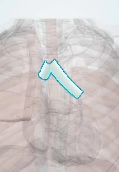
Structures involved
Tap one to see what it is and open it in 3D.
In biochemistry
Microorganisms that cause it
Why it happens
- Special stains in diagnosis: Congo red, Prussian blue, PAS, Ziehl–Neelsen, Gram
- Diffusion across the respiratory membrane and DLCO
- The causes of hypoxemia
- The alveolus: type I and II pneumocytes, surfactant, the blood–air barrier
What it is
An opportunistic pneumonia caused by the fungus Pneumocystis jirovecii in people with impaired cell-mediated immunity. It is a classic AIDS-defining illness and also affects transplant recipients, people with cancer and those on long-term corticosteroids.
Who gets it
Remains one of the most frequent AIDS-defining illnesses in Latin America, often the event that reveals a late HIV diagnosis. Its share of cases from non-HIV immunosuppression (transplants, chemotherapy, biologics, corticosteroids) is growing.
Causes
- Pneumocystis jirovecii, an atypical fungus that cannot be cultured routinely
- Airborne transmission between people; healthy carriers are common
- Disease occurs when T-cell immunity falls
Risk factors
- HIV with CD4 count below 200 cells/µL or CD4 below 14%
- Long-term high-dose corticosteroids, especially with another immunosuppressant
- Solid organ or haematopoietic stem cell transplant
- Haematological cancers and chemotherapy
- Not receiving prophylaxis when indicated
Pathophysiology
Inhaled organisms attach to type I pneumocytes and multiply in the alveoli. A foamy exudate of organisms and proteinaceous material fills the alveoli, causing interstitial inflammation, impaired gas exchange and hypoxaemia that is out of proportion to the findings on examination. The host inflammatory response worsens lung injury, especially after treatment begins, which is why corticosteroids help in hypoxaemic patients.
Classification
- Mild: PaO2 ≥ 70 mmHg on room air
- Moderate to severe: PaO2 below 70 mmHg or alveolar-arterial gradient ≥ 35 mmHg
Symptoms and signs
- Gradually worsening shortness of breath on exertion over days to weeks (subacute in HIV)
- Dry, non-productive cough
- Fever and fatigue
- Tachypnoea and desaturation on exertion with few findings on chest auscultation
- Faster and more severe course in people without HIV
Diagnosis
- Clinical
- Subacute dyspnoea, dry cough and fever in a person with known or suspected HIV or other immunosuppression; ask about prophylaxis and CD4 count. Exercise desaturation is a useful bedside clue.
- Tests
- Arterial blood gases (hypoxaemia, widened A-a gradient), raised LDH, serum beta-D-glucan. Confirmation by detecting the organism in induced sputum or bronchoalveolar lavage with immunofluorescence, silver (methenamine) stains or PCR. HIV test and CD4 count.
- Imaging
- Chest X-ray: bilateral, symmetric perihilar interstitial infiltrates, sometimes normal early. CT shows bilateral ground-glass opacities and may reveal cysts (pneumatoceles) that predispose to pneumothorax.
Differential diagnosis
- Pulmonary tuberculosis
- Bacterial community-acquired pneumonia
- Cytomegalovirus pneumonitis
- Histoplasmosis and other endemic mycoses
- Pulmonary Kaposi sarcoma
Goal of treatment
Eradicate the infection, correct hypoxaemia and limit the inflammatory lung injury; then prevent relapse with secondary prophylaxis and immune recovery (antiretroviral therapy in HIV).
Treatment
- Medical
- Trimethoprim-sulfamethoxazole is first-line treatment, orally or intravenously according to severity; alternatives for intolerance include primaquine with clindamycin, atovaquone and pentamidine.
- Medical
- Adjunctive corticosteroids for moderate to severe disease with hypoxaemia, started early alongside antimicrobial treatment.
- Acute
- Supplemental oxygen and, in respiratory failure, non-invasive or mechanical ventilation in intensive care.
- Medical
- Start antiretroviral therapy within the first weeks of treatment in people with HIV, watching for immune reconstitution inflammatory syndrome.
- Follow-up
- Monitoring of oxygen saturation and respiratory effort, drug toxicity (rash, cytopenias, hyperkalaemia, kidney function) and continuation of secondary prophylaxis until CD4 recovers.
Drugs for this condition
Complications
- Respiratory failure and acute respiratory distress syndrome
- Pneumothorax from cystic lesions
- Adverse reactions to sulfonamides (rash, Stevens-Johnson syndrome, cytopenias)
- Immune reconstitution inflammatory syndrome
- Death, especially when diagnosis or treatment is delayed
Prognosis
With early treatment most people with HIV survive, but mortality rises sharply with respiratory failure and is higher in non-HIV immunosuppressed patients. Relapse is common without prophylaxis or immune recovery.
Prevention
- Primary prophylaxis with trimethoprim-sulfamethoxazole in HIV with CD4 below 200 cells/µL and in transplant or high-risk immunosuppressed patients
- Early HIV testing and antiretroviral therapy
- Secondary prophylaxis after an episode until immune recovery
Test yourself
Think of the answer, then tap the question to see it.
Below what CD4 count is prophylaxis against Pneumocystis started in HIV?
Below 200 cells/µL (or CD4 below 14%).
What is the classic chest X-ray of Pneumocystis pneumonia?
Bilateral, symmetric perihilar interstitial (ground-glass) infiltrates; CT shows ground-glass opacities and sometimes cysts.
What is the first-line drug for treatment and prophylaxis?
Trimethoprim-sulfamethoxazole.
When are corticosteroids added to treatment?
In moderate to severe disease with hypoxaemia (PaO2 below 70 mmHg or A-a gradient of 35 mmHg or more).
Which stain shows Pneumocystis cysts in lavage fluid?
Silver (methenamine silver) stain, or direct immunofluorescence.
Related conditions
- HIV infection and AIDS
- Community-acquired pneumonia
- Pulmonary tuberculosis
- Acute respiratory distress syndrome (ARDS)
- Histoplasmosis
- Cytomegalovirus infection
- Kaposi sarcoma
- Toxoplasmosis
- Cryptococcal meningitis
Educational information, not medical advice. For any symptom, see a health professional.
Sources: Pneumocystis Pneumonia Basics (Centers for Disease Control and Prevention (CDC), 2024); Pneumocystis jirovecii pneumonia: MedlinePlus Medical Encyclopedia (MedlinePlus (U.S. National Library of Medicine), 2024).
Spotted a mistake?We are always improving for the community. Tell us what is wrong and our team will review it.Report a mistake
Representative model, for educational purposes only.