Conditions · Pediatrics and neonatology
Posterior urethral valves !
Posterior urethral valves are folds of tissue in a baby boy's urethra that block urine flow and can damage the bladder and kidneys.
Updated 2026-11-01
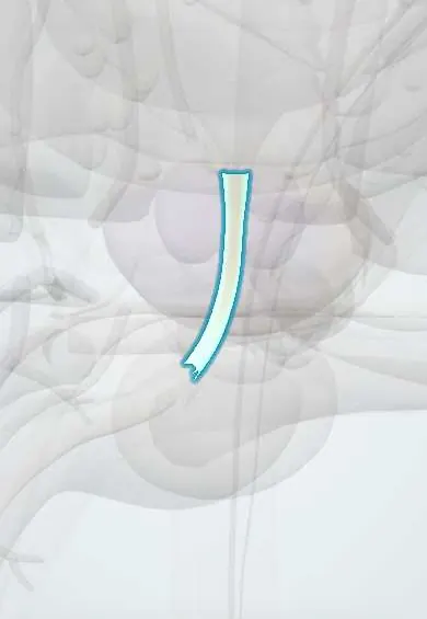
Structures involved
Tap one to see what it is and open it in 3D.
Why it happens
- Hypertrophy: physiologic and pathologic
- Micturition and the bladder reflex
- Ureter, bladder and urethra: urothelium
What it is
A congenital obstruction of the male posterior (prostatic) urethra caused by abnormal membranous folds. It is the most common cause of lower urinary tract obstruction in boys and a leading congenital cause of chronic kidney disease in children.
Who gets it
Occurs only in males, in roughly 1 in 5,000 to 8,000 live-born boys. Most cases are now suspected on prenatal ultrasound; in settings with limited prenatal screening, as in parts of Latin America, diagnosis may come later, after urinary infection, poor growth or kidney failure.
Causes
- Abnormal development of the posterior urethra early in fetal life, probably from faulty insertion of the Wolffian (mesonephric) duct remnants
- Usually sporadic; a familial pattern is rare
Risk factors
- Male sex (the only group affected)
- Prenatal finding of bilateral hydronephrosis with a thick-walled, distended bladder
Pathophysiology
The valves act as a partial dam below the bladder neck. The bladder must generate high pressure to empty, so its wall thickens and becomes trabeculated and poorly compliant. High pressure is transmitted up the ureters, causing hydroureteronephrosis, often vesicoureteral reflux, and damage to the developing kidneys (renal dysplasia). Because fetal urine makes most of the amniotic fluid after mid-pregnancy, severe obstruction causes oligohydramnios, which compresses the fetus and impairs lung growth (pulmonary hypoplasia, Potter sequence).
Classification
- Young type I: folds extending distally from the verumontanum (most common)
- Young type III: a membrane with a central opening (ring or diaphragm) below the verumontanum
- Young type II (folds toward the bladder neck) is no longer considered obstructive
Symptoms and signs
- Prenatal: bilateral hydronephrosis, distended thick-walled bladder with dilated posterior urethra (keyhole sign), oligohydramnios
- Newborn: palpable bladder, weak or dribbling urine stream, straining to void
- Respiratory distress from pulmonary hypoplasia in severe cases
- Older infants: urinary tract infection, sepsis, poor feeding and failure to thrive
- Older boys: daytime incontinence, frequency or urgency, poor stream
Diagnosis
- Clinical
- Suspected in a boy with prenatal bilateral hydronephrosis and a full bladder, or in a newborn or infant boy with a poor stream, urinary infection or kidney dysfunction.
- Tests
- Serum creatinine (trend after the first days of life, since newborn values reflect the mother's), electrolytes and acid-base status, urinalysis and urine culture. Cystoscopy both confirms and treats. Urodynamic studies assess bladder function at follow-up.
- Imaging
- Voiding cystourethrogram (VCUG) is the key test: dilated, elongated posterior urethra with an abrupt narrowing at the valve, a trabeculated bladder and often reflux. Renal and bladder ultrasound shows hydronephrosis, a thick-walled bladder and signs of dysplasia.
Differential diagnosis
- Prune belly syndrome
- Bilateral ureteropelvic or ureterovesical junction obstruction
- High-grade vesicoureteral reflux without obstruction
- Neurogenic bladder (for example with spina bifida)
- Urethral atresia or stricture
Goal of treatment
Relieve the obstruction early, protect kidney function and manage the bladder for life to delay or prevent kidney failure.
Treatment
- Acute
- At birth: bladder drainage with a urethral catheter, correction of fluid, electrolyte and acid-base problems, and respiratory support if needed.
- Surgical
- Endoscopic valve ablation (cystoscopic incision of the valves) once the baby is stable; a temporary vesicostomy if the urethra is too small.
- Medical
- Antibiotic treatment of urinary infections and, in selected children, prophylaxis; anticholinergic drugs for an overactive bladder; management of chronic kidney disease.
- Follow-up
- Lifelong follow-up of kidney function, blood pressure, growth and bladder function (valve bladder), with clean intermittent catheterization when the bladder empties poorly.
Complications
- Chronic kidney disease and kidney failure needing dialysis or transplant
- Pulmonary hypoplasia and neonatal death in severe cases
- Recurrent urinary tract infection and urosepsis
- Valve bladder syndrome: poorly compliant, poorly emptying bladder with incontinence
- Polyuria from tubular damage, with risk of dehydration
Prognosis
Depends on the degree of kidney damage before relief. A low creatinine nadir in the first year of life predicts better long-term kidney function; a substantial minority (up to about a quarter or more) reach kidney failure over their lifetime.
Prevention
- No known prevention; prenatal ultrasound allows planned delivery and early drainage and ablation
Test yourself
Think of the answer, then tap the question to see it.
What is the most common cause of congenital lower urinary tract obstruction in boys?
Posterior urethral valves.
Which study is the key test to diagnose posterior urethral valves?
Voiding cystourethrogram, showing a dilated, elongated posterior urethra with abrupt narrowing at the valves.
Why can posterior urethral valves cause respiratory distress at birth?
Severe obstruction reduces fetal urine and amniotic fluid; oligohydramnios causes pulmonary hypoplasia (Potter sequence).
What is the definitive first-line treatment once the newborn is stable?
Endoscopic (cystoscopic) valve ablation, after initial bladder drainage with a catheter.
Which laboratory marker in the first year predicts long-term kidney outcome?
The lowest (nadir) serum creatinine after relief of obstruction.
Related conditions
- Hydronephrosis and obstructive uropathy
- Vesicoureteral reflux
- Chronic kidney disease
- Urinary tract infection (acute cystitis)
- Congenital kidney anomalies (horseshoe kidney, renal agenesis)
- Neurogenic bladder
- Acute kidney injury
- Hyperkalaemia
Educational information, not medical advice. For any symptom, see a health professional.
Sources: Posterior Urethral Valves (Boston Children's Hospital, 2024); Potter syndrome (MedlinePlus (U.S. National Library of Medicine), 2024).
Spotted a mistake?We are always improving for the community. Tell us what is wrong and our team will review it.Report a mistake
Representative model, for educational purposes only.