Conditions · Nephrology and urology
Congenital kidney anomalies (horseshoe kidney, renal agenesis) !
Kidneys and ureters that form in the wrong number, place or shape before birth. Many cause no symptoms; some block urine or limit kidney function.
Updated 2026-11-01
Structures involved
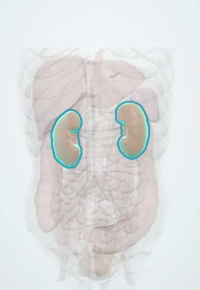
Tap one to see what it is and open it in 3D.
Why it happens
What it is
A group of birth defects of the kidneys and urinary tract (often called CAKUT) that includes missing kidneys (agenesis), fused kidneys (horseshoe kidney), kidneys in the wrong place (ectopic or pelvic kidney), small or abnormal kidneys and obstruction where the renal pelvis meets the ureter. They are the leading cause of chronic kidney disease in children.
Who gets it
Together they are among the most common birth defects found on prenatal ultrasound. Horseshoe kidney occurs in roughly 1 in 400–600 people and is more common in males; unilateral renal agenesis in roughly 1 in 1,000–2,000; bilateral agenesis is rare and incompatible with life. In Colombia, most are detected on routine prenatal ultrasound during antenatal care.
Causes
- Failure of the ureteric bud to reach or induce the metanephric mesenchyme (renal agenesis)
- Fusion of the lower poles of both kidneys, which then become trapped under the inferior mesenteric artery during ascent (horseshoe kidney)
- Failure of the kidney to ascend from the pelvis (pelvic or ectopic kidney)
- Narrowing or abnormal muscle at the ureteropelvic junction, or a crossing lower-pole vessel (ureteropelvic junction obstruction)
- Genetic syndromes and gene variants that affect kidney development
Risk factors
- Family history of kidney or urinary tract malformations
- Chromosomal syndromes: Turner syndrome (horseshoe kidney), trisomies 13, 18 and 21
- Maternal diabetes during pregnancy
- Other birth defects, for example of the genital tract, spine, heart or anorectum (VACTERL association)
Pathophysiology
The permanent kidney (metanephros) forms when the ureteric bud grows into the metanephric mesenchyme and each induces the other. If this signalling fails, no kidney forms. The kidneys then ascend from the pelvis to the lumbar region; fused lower poles catch on the inferior mesenteric artery and stay low as a horseshoe kidney, whose ureters drain poorly, favouring stasis, stones and infection. With bilateral agenesis the fetus makes no urine, so amniotic fluid is lacking (oligohydramnios) and the lungs fail to develop: Potter sequence, with flat face, limb deformities and fatal pulmonary hypoplasia. A single kidney grows larger (compensatory hypertrophy) and its nephrons work harder, which over decades can lead to hypertension and proteinuria.
Classification
- Anomalies of number: unilateral or bilateral renal agenesis, supernumerary kidney
- Anomalies of position and fusion: pelvic or ectopic kidney, crossed renal ectopia, horseshoe kidney
- Anomalies of the kidney tissue: hypoplasia, dysplasia, multicystic dysplastic kidney
- Anomalies of the collecting system: ureteropelvic junction obstruction, duplicated ureter, ureterocele, vesicoureteral reflux
Symptoms and signs
- Often none; found by chance on prenatal or abdominal ultrasound
- Recurrent urinary tract infections
- Flank or abdominal pain, sometimes after drinking large volumes (ureteropelvic junction obstruction)
- A palpable abdominal mass in an infant (hydronephrosis or multicystic kidney)
- Kidney stones or blood in the urine
- High blood pressure or protein in the urine in later life with a single kidney
Diagnosis
- Clinical
- Suspect it from a prenatal ultrasound finding, recurrent urinary infections in a child, a neonatal abdominal mass or an associated syndrome such as Turner syndrome.
- Tests
- Urinalysis and urine culture, creatinine with estimated GFR, urine albumin or protein and blood pressure measurement; genetic testing when a syndrome is suspected.
- Imaging
- Kidney and bladder ultrasound is the first test (prenatal and postnatal). Voiding cystourethrography looks for reflux, MAG3 diuretic renography measures drainage and split function in suspected obstruction, and CT or MR urography defines complex anatomy.
Differential diagnosis
- Prior nephrectomy or an atrophic kidney after injury
- Autosomal recessive or dominant polycystic kidney disease
- Posterior urethral valves
- Wilms tumour or neuroblastoma (abdominal mass in a child)
- Non-obstructive dilatation of the renal pelvis
Goal of treatment
Preserve kidney function, prevent infection and stones, relieve true obstruction and screen for associated anomalies.
Treatment
- Follow-up
- Follow-up ultrasound after birth for prenatal findings, and lifelong checks of blood pressure, urine protein and kidney function with a single kidney.
- Conservative
- Most fusion and position anomalies need no treatment; avoid nephrotoxic drugs and dehydration, and protect the kidney from injury in contact sports per individual advice.
- Surgical
- Pyeloplasty (open, laparoscopic or robotic) for ureteropelvic junction obstruction with symptoms, worsening dilatation or falling split function.
- Medical
- Treatment of urinary infections and, in selected children with reflux, antibiotic prophylaxis; blood-pressure control and renin–angiotensin blockers for proteinuria.
- Acute
- Neonatal intensive care for a newborn with suspected Potter sequence; prompt antibiotic treatment of febrile infections.
Complications
- Hydronephrosis and loss of kidney function from obstruction
- Recurrent pyelonephritis and kidney scarring
- Kidney stones (especially in horseshoe kidney)
- Hypertension, proteinuria and chronic kidney disease
- Pulmonary hypoplasia and neonatal death in bilateral agenesis
- Higher risk of injury of a horseshoe kidney in abdominal trauma; a slightly higher rate of some kidney tumours, including Wilms tumour
Prognosis
Most people with a single kidney or horseshoe kidney live normal lives with periodic monitoring. Outcome depends on how much functional kidney tissue there is and on associated anomalies; bilateral agenesis is fatal.
Prevention
- Good prenatal care with routine ultrasound and control of maternal diabetes
- Early detection and follow-up to protect the remaining kidney function
Test yourself
Think of the answer, then tap the question to see it.
Which artery traps the ascending horseshoe kidney?
The inferior mesenteric artery.
Which syndrome is classically associated with horseshoe kidney?
Turner syndrome (45,X).
What are the features of Potter sequence and its cause of death?
Oligohydramnios from absent fetal urine (e.g. bilateral renal agenesis) causes a flat face, limb deformities and pulmonary hypoplasia, which is the cause of death.
What is the most common obstructive cause of hydronephrosis detected in newborns?
Ureteropelvic junction obstruction (transient, non-obstructive dilatation is even more frequent overall).
Related conditions
- Vesicoureteral reflux
- Hydronephrosis and obstructive uropathy
- Posterior urethral valves
- Turner syndrome
- Wilms tumour (nephroblastoma)
- Kidney stones (urolithiasis)
- Urinary tract infection (acute cystitis)
- Acute pyelonephritis
- Chronic kidney disease
- Autosomal dominant polycystic kidney disease
Educational information, not medical advice. For any symptom, see a health professional.
Sources: Solitary or Single-functioning Kidney - NIDDK (NIDDK (National Institutes of Health), 2024); Ectopic Kidney - NIDDK (NIDDK (National Institutes of Health), 2024).
Spotted a mistake?We are always improving for the community. Tell us what is wrong and our team will review it.Report a mistake
Representative model, for educational purposes only.