Conditions · Nephrology and urology
Vesicoureteral reflux !
Urine flows backward from the bladder up the ureters toward the kidneys. In children it raises the risk of kidney infections and scarring.
Updated 2026-11-01
Structures involved
Tap one to see what it is and open it in 3D.
In embryology
- Kidney: pronephros, mesonephros, metanephros and ascent
- Bladder, urachus and urethra
- Horseshoe kidney and renal ectopia
- Duplicated ureter, UPJ obstruction and posterior urethral valves
- Bladder exstrophy
Why it happens
What it is
Retrograde flow of urine from the bladder into one or both ureters and sometimes the kidneys, usually because the valve-like junction where the ureter enters the bladder does not close properly. It is mainly a disease of infants and young children.
Who gets it
Found in a large share of young children investigated after a febrile urinary infection, and in some siblings and children of affected people. Many cases are detected after a first febrile urinary infection or because of hydronephrosis on prenatal ultrasound.
Causes
- Primary: a short intramural (submucosal) ureter segment at the ureterovesical junction, which tends to lengthen with growth
- Secondary: high bladder pressure from posterior urethral valves, neurogenic bladder or bladder–bowel dysfunction
- Familial tendency
Risk factors
- Sibling or parent with reflux
- Infancy and young age
- Hydronephrosis detected before birth
- Bladder–bowel dysfunction (holding urine, constipation)
- Posterior urethral valves or neurogenic bladder
Pathophysiology
The ureter normally tunnels obliquely through the bladder wall, so bladder filling compresses it shut. A short or laterally placed tunnel lets urine flow back during filling or voiding. Bacteria in the bladder then reach the kidney, causing pyelonephritis, and repeated infection in a growing kidney leads to scarring (reflux nephropathy).
Classification
- International grading I–V on voiding cystourethrography: I = ureter only; II = up to the pelvis without dilatation; III–IV = increasing dilatation of ureter and calyces; V = severe tortuous dilatation with loss of papillary impressions
- Primary or secondary
- Unilateral or bilateral
Symptoms and signs
- Often none until a urinary infection occurs
- Febrile urinary tract infection in infants: fever, irritability, poor feeding, vomiting
- In older children: painful or frequent urination, flank or abdominal pain
- Daytime wetting or constipation (bladder–bowel dysfunction)
- Hydronephrosis found on prenatal ultrasound
Diagnosis
- Clinical
- Suspect it in infants and children with febrile urinary infection, a family history or prenatal hydronephrosis; ask about voiding and bowel habits and check blood pressure and growth.
- Tests
- Urinalysis and urine culture from a properly collected sample; creatinine and urine protein when there is scarring.
- Imaging
- Kidney and bladder ultrasound first. Voiding cystourethrography (VCUG) is the reference test to diagnose and grade reflux; DMSA scan detects kidney scarring.
Differential diagnosis
- Ureteropelvic junction obstruction
- Posterior urethral valves
- Primary megaureter or ureterocele
- Neurogenic bladder
- Recurrent cystitis without reflux
Goal of treatment
Prevent febrile urinary infections and kidney scarring while waiting for spontaneous resolution, and correct reflux when it persists or causes harm.
Treatment
- Follow-up
- Watchful waiting with prompt urine testing and treatment of every febrile illness; low grades often resolve with growth.
- Medical
- Prompt antibiotic treatment of infections; continuous low-dose antibiotic prophylaxis in selected children, especially higher grades or recurrent infections.
- Lifestyle
- Treating bladder–bowel dysfunction: regular voiding, good fluid intake and correcting constipation; circumcision may be discussed in boys with high-grade reflux.
- Procedure
- Endoscopic injection of a bulking agent at the ureteral opening.
- Surgical
- Ureteral reimplantation (open, laparoscopic or robotic) for breakthrough infections, new scarring or persistent high-grade reflux; secondary reflux is treated at its cause.
Complications
- Recurrent pyelonephritis and urosepsis
- Kidney scarring (reflux nephropathy)
- Hypertension
- Chronic kidney disease and, rarely, kidney failure
- Pregnancy complications in women with scarring
Prognosis
Most low-grade primary reflux resolves within a few years as the child grows. High-grade and bilateral reflux resolves less often and carries the highest risk of scarring, hypertension and kidney disease.
Prevention
- Early diagnosis and treatment of febrile urinary infections in infants
- Screening of siblings in selected families
Test yourself
Think of the answer, then tap the question to see it.
What is the reference imaging test to diagnose and grade vesicoureteral reflux?
Voiding cystourethrography (VCUG).
What causes primary vesicoureteral reflux?
A short intravesical (submucosal) ureteral tunnel at the ureterovesical junction, so the valve does not close.
Which test detects kidney scarring?
A DMSA renal scan.
What is the main long-term risk of high-grade reflux?
Reflux nephropathy: kidney scarring leading to hypertension and chronic kidney disease.
Related conditions
- Urinary tract infection (acute cystitis)
- Acute pyelonephritis
- Hydronephrosis and obstructive uropathy
- Posterior urethral valves
- Neurogenic bladder
- Chronic kidney disease
- Arterial hypertension
- Spina bifida (myelomeningocele)
Educational information, not medical advice. For any symptom, see a health professional.
Sources: Vesicoureteral Reflux (VUR) (NIDDK (National Institutes of Health), 2024); Bilateral hydronephrosis: MedlinePlus Medical Encyclopedia (MedlinePlus (U.S. National Library of Medicine), 2024).
Spotted a mistake?We are always improving for the community. Tell us what is wrong and our team will review it.Report a mistake
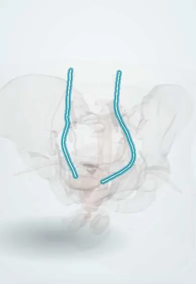
Representative model, for educational purposes only.