Conditions · Nephrology and urology
Neurogenic bladder !
Bladder control problems caused by damage to the brain, spinal cord or nerves. The bladder may empty too often, too little, or not at all.
Updated 2026-11-01
Structures involved
Tap one to see what it is and open it in 3D.
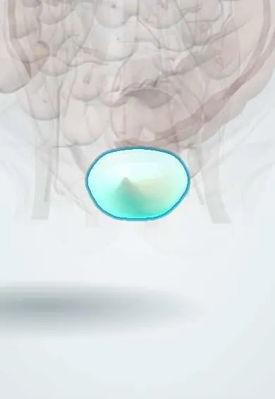
- Urinary bladder
- Conus medullaris
- External urethral sphincter
- Spinal cord
- Cauda equina
- Pudendal nerve
- Ureters
- Trigone of bladder
- Kidneys
In embryology
- Spinal cord: alar and basal plates and the ascent of the cord
- Neural tube defects: anencephaly, spina bifida and encephalocele
- Congenital Zika syndrome
In the clinical exam
Why it happens
- Micturition and the bladder reflex
- Upper versus lower motor neuron lesions
- The autonomic nervous system: organization
What it is
Bladder and sphincter dysfunction caused by disease of the nervous system controlling urination. Depending on the level of the lesion, the bladder may be overactive (spastic) or underactive (flaccid), and the bladder and sphincter may fail to coordinate.
Who gets it
Very common after spinal cord injury and in spina bifida, multiple sclerosis, stroke and Parkinson disease. In Colombia, traffic and violence-related spinal cord injuries and myelomeningocele in children make it a frequent problem in rehabilitation and paediatric urology.
Causes
- Spinal cord injury
- Spina bifida (myelomeningocele) and tethered cord
- Multiple sclerosis and transverse myelitis
- Stroke, Parkinson disease and dementia (lesions above the pons)
- Diabetic autonomic neuropathy
- Cauda equina syndrome, lumbar disc disease and pelvic surgery damaging the pelvic nerves
Risk factors
- Spinal cord injury or congenital spinal defects
- Long-standing diabetes
- Progressive neurological disease
- Radical pelvic surgery
Pathophysiology
Voiding is coordinated by the pontine micturition centre, with sacral (S2–S4) parasympathetic fibres contracting the detrusor and pudendal fibres controlling the external sphincter. Lesions above the pons give an overactive detrusor with coordinated voiding. Spinal lesions between the pons and the sacral cord give an overactive detrusor with detrusor–sphincter dyssynergia and high bladder pressures. Lesions of the conus, cauda equina or peripheral nerves give an underactive, large bladder with retention and overflow.
Classification
- Suprapontine lesion: urge incontinence, coordinated sphincter, low residual
- Suprasacral spinal lesion: spastic bladder with detrusor–sphincter dyssynergia
- Sacral or infrasacral lesion (conus, cauda equina, peripheral nerve): flaccid bladder with retention and overflow
Symptoms and signs
- Urgency, frequency and urge incontinence (overactive type)
- Difficulty starting, weak stream, incomplete emptying (underactive type)
- Overflow leakage with a full bladder and no sensation of fullness
- Recurrent urinary infections
- Associated bowel dysfunction and sexual dysfunction
Diagnosis
- Clinical
- Neurological history and examination (sensation, reflexes, saddle area, anal tone), bladder diary and voiding pattern. Urodynamic studies define the type and identify high-pressure bladders.
- Tests
- Post-void residual volume, urinalysis and culture when infection is suspected, creatinine. Urodynamics with pressure–flow studies (and electromyography of the sphincter) is the key test.
- Imaging
- Kidney and bladder ultrasound for hydronephrosis, residual and stones; voiding cystourethrography for reflux; spinal MRI to look for the neurological cause.
Differential diagnosis
- Benign prostatic hyperplasia or urethral stricture
- Idiopathic overactive bladder
- Stress urinary incontinence
- Drug-induced retention (anticholinergics, opioids)
- Pelvic organ prolapse
Goal of treatment
Protect the upper urinary tract (keep bladder pressures low), achieve regular complete emptying and continence, and prevent infection.
Treatment
- Procedure
- Clean intermittent catheterisation on a schedule is the standard for incomplete emptying; teaching the patient or carer is a key nursing task. Indwelling catheters are avoided where possible.
- Medical
- Antimuscarinic drugs or beta-3 agonists to lower detrusor pressure in overactive bladders; alpha-blockers may help outlet resistance in selected cases.
- Rehabilitation
- Bladder and bowel programmes, timed voiding, pelvic floor work and adequate fluid intake.
- Surgical
- For refractory cases: botulinum toxin into the detrusor, bladder augmentation, urinary diversion or sacral neuromodulation.
- Follow-up
- Lifelong follow-up with kidney ultrasound, creatinine and periodic urodynamics; monitoring for infection, stones and autonomic dysreflexia.
Drugs for this condition
Complications
- Recurrent urinary infection and pyelonephritis
- Vesicoureteral reflux, hydronephrosis and chronic kidney disease
- Bladder stones
- Autonomic dysreflexia in high spinal cord injury
- Skin breakdown from leakage
Prognosis
A long-term condition. With intermittent catheterisation and regular follow-up most people keep good kidney function; kidney failure from high bladder pressures, once a common cause of death after spinal injury, is now largely preventable.
Prevention
- Folic acid before and in early pregnancy to reduce neural tube defects
- Early urological assessment after spinal cord injury and in spina bifida
Test yourself
Think of the answer, then tap the question to see it.
What is detrusor–sphincter dyssynergia and which lesion causes it?
The detrusor contracts while the external sphincter fails to relax, typically with spinal cord lesions between the pons and the sacral cord.
What type of bladder results from a conus or cauda equina lesion?
A flaccid, underactive bladder with retention and overflow incontinence.
What is the standard method to empty the bladder in neurogenic retention?
Clean intermittent catheterisation on a regular schedule.
What is the most common trigger of autonomic dysreflexia?
A distended bladder (blocked catheter, retention), followed by constipation.
Related conditions
- Spinal cord injury
- Spina bifida (myelomeningocele)
- Multiple sclerosis
- Parkinson disease
- Ischaemic stroke
- Peripheral neuropathy (diabetic and other)
- Cauda equina syndrome
- Autonomic dysreflexia
- Urinary tract infection (acute cystitis)
- Hydronephrosis and obstructive uropathy
- Vesicoureteral reflux
- Overactive bladder and urge incontinence
Educational information, not medical advice. For any symptom, see a health professional.
Sources: Neurogenic bladder: MedlinePlus Medical Encyclopedia (MedlinePlus (U.S. National Library of Medicine), 2024).
Spotted a mistake?We are always improving for the community. Tell us what is wrong and our team will review it.Report a mistake
Representative model, for educational purposes only.