Conditions · Infectious and tropical diseases
Q fever !
A zoonosis caught by breathing dust from goats, sheep or cattle; it causes flu-like illness, pneumonia or hepatitis, and rarely a chronic heart-valve infection.
Updated 2026-11-01
Structures involved
Tap one to see what it is and open it in 3D.
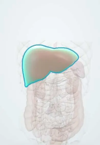
- Liver
- Gallbladder
- Spleen
- Portal vein
- Pleura
- Aortic right coronary cusp
- Aortic non-coronary cusp
- Mitral anterior leaflet
- Pericardium
Microorganisms that cause it
Why it happens
- Granulomatous inflammation: caseating vs non-caseating, the causes to know
- Liver: lobule, acinus, hepatocytes, sinusoids, Kupffer cells
- The heart wall, valves and conduction tissue
What it is
Infection by Coxiella burnetii, a small intracellular bacterium carried by farm animals. Most infections are silent or self-limited, but a minority cause acute pneumonia or hepatitis, and a small group, mostly people with damaged heart valves or vascular grafts, develops persistent (chronic) infection, classically culture-negative endocarditis.
Who gets it
Found worldwide except New Zealand. Outbreaks follow contact with lambing or calving animals, such as the large Dutch goat-farm epidemic of 2007–2010. In Colombia and the rest of Latin America, serological studies show exposure in cattle, goats and rural workers, but the disease is rarely tested for and is probably underdiagnosed among causes of fever with pneumonia or hepatitis.
Causes
- Coxiella burnetii, a Gram-negative obligate intracellular bacterium that forms a resistant spore-like form surviving months in dust
- Main reservoirs: goats, sheep and cattle; also cats, dogs and wild animals; ticks maintain it in nature
- Inhalation of aerosols from placenta, birth fluids, feces, urine or contaminated dust; wind can carry it kilometres
- Less often: raw milk and unpasteurized dairy products; person-to-person spread is exceptional
Risk factors
- Work with livestock: farmers, ranchers, dairy and slaughterhouse workers, veterinarians and laboratory staff
- Contact with animals giving birth or with their placenta
- Drinking raw milk
- For chronic infection: heart valve disease or prosthetic valve, aortic aneurysm or vascular graft, immunosuppression and pregnancy
Pathophysiology
Inhaled bacteria are taken up by alveolar macrophages and survive inside an acidic phagolysosome, where they multiply. They spread through the blood to the liver, spleen, bone marrow and other organs, where the immune response forms granulomas; in the liver these are characteristically ring-shaped («doughnut» granulomas). When cell-mediated immunity fails to clear the organism, it persists on abnormal valves or vascular endothelium. The bacterium shows antigenic phase variation: antibodies to phase II antigen predominate in acute infection, while high phase I IgG points to persistent infection.
Classification
- Asymptomatic seroconversion (about half of infections)
- Acute Q fever: flu-like febrile illness, atypical pneumonia or granulomatous hepatitis
- Persistent (chronic) Q fever: endocarditis, vascular graft or aneurysm infection, osteomyelitis
- Post-Q fever fatigue syndrome
Symptoms and signs
- Abrupt high fever, chills and an intense headache, often behind the eyes, two to three weeks after exposure
- Muscle aches, sweats and marked fatigue
- Dry cough and pleuritic chest pain when the lung is involved
- Nausea, abdominal pain and sometimes jaundice when the liver is involved
- In chronic infection: long-lasting low fever, weight loss, night sweats, new heart murmur and heart failure
Diagnosis
- Clinical
- Suspected in fever with pneumonia or hepatitis after contact with livestock, birth products or raw milk. Routine blood cultures do not grow the organism, so it must be requested specifically.
- Tests
- Serology by indirect immunofluorescence is the standard: a fourfold rise in phase II IgG between acute and convalescent samples confirms acute infection, and a high phase I IgG titre suggests persistent infection. PCR on blood or serum is useful in the first two weeks and on valve or tissue samples. Laboratory clues: raised liver enzymes, normal or low white count and low platelets.
- Imaging
- Chest X-ray shows segmental or rounded opacities that are not specific. Transesophageal echocardiography looks for vegetations, which are often small or absent; PET-CT helps find vascular or graft infection.
Differential diagnosis
- Other atypical pneumonias (Mycoplasma, Legionella, psittacosis)
- Brucellosis and leptospirosis
- Viral hepatitis and other granulomatous hepatitis
- Rickettsial infections, dengue and influenza
- Other causes of culture-negative endocarditis (Bartonella, prior antibiotics)
Goal of treatment
Shorten acute illness, cure persistent infection with prolonged combined antibiotics and protect high-risk patients (valve disease, pregnancy) from chronic infection.
Treatment
- Medical
- Acute symptomatic Q fever: a tetracycline (doxycycline) for about two weeks; other options in pregnancy and young children are chosen by specialists.
- Medical
- Persistent infection or endocarditis: doxycycline combined with hydroxychloroquine, which makes the drug bactericidal inside the acidic vacuole, for at least 18 months (longer with prosthetic material).
- Surgical
- Valve replacement or removal of an infected vascular graft when there is heart failure, a large vegetation or no response to antibiotics.
- Follow-up
- Serial serology (falling phase I antibodies) to judge cure; eye checks during hydroxychloroquine and sun protection during doxycycline; echocardiographic screening of valve disease after acute infection.
Complications
- Endocarditis, often on previously damaged or prosthetic valves
- Infection of aortic aneurysms and vascular grafts
- Myocarditis and pericarditis
- Miscarriage, preterm birth and fetal death
- Osteomyelitis and chronic fatigue after infection
Prognosis
Acute Q fever is usually self-limited, with mortality below 2%. Persistent infection is serious: endocarditis and vascular infection carry significant mortality even with treatment, and need long therapy and follow-up.
Prevention
- Pasteurization of milk and avoidance of raw dairy products
- Safe handling and disposal of placentas and birth products; protective equipment for animal workers
- Vaccination of herds and, where available, of high-risk workers
- Counselling people with valve disease or pregnancy to avoid exposure
Test yourself
Think of the answer, then tap the question to see it.
What is the main route of infection in Q fever?
Inhalation of dust or aerosols contaminated by birth products, urine or feces of goats, sheep or cattle.
What is the classic presentation of chronic Q fever?
Culture-negative endocarditis, usually in a patient with prior valve disease or a prosthetic valve.
Which serological pattern suggests persistent infection?
A high IgG titre against phase I antigen (acute infection is dominated by phase II antibodies).
What is the treatment for Q fever endocarditis?
Doxycycline plus hydroxychloroquine for at least 18 months, with surgery when needed.
What liver lesion is typical of acute Q fever?
Granulomatous hepatitis with ring-shaped («doughnut») granulomas.
Related conditions
- Infective endocarditis
- Community-acquired pneumonia
- Viral hepatitis
- Brucellosis
- Leptospirosis
- Rickettsial infections (spotted fever)
- Psittacosis
- Legionnaires disease and Pontiac fever
- Miscarriage (early pregnancy loss)
- Osteomyelitis
Educational information, not medical advice. For any symptom, see a health professional.
Sources: About Q fever | Q Fever | CDC (Centers for Disease Control and Prevention, 2024); Q fever: MedlinePlus Medical Encyclopedia (MedlinePlus (U.S. National Library of Medicine), 2024).
Spotted a mistake?We are always improving for the community. Tell us what is wrong and our team will review it.Report a mistake
Representative model, for educational purposes only.