Chagas cardiomyopathy !
Chagas cardiomyopathy is heart disease caused by the parasite Trypanosoma cruzi, appearing years after infection with arrhythmias and heart failure.
Updated 2026-10-29
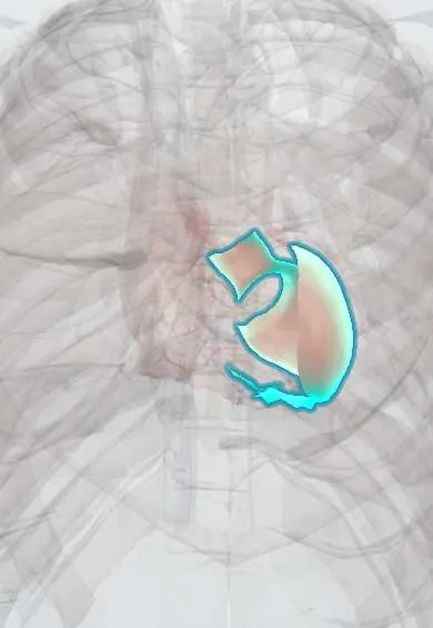
Structures involved
Tap one to see what it is and open it in 3D.
- Left ventricle
- Bundle branches
- Right ventricle
- Atrioventricular (AV) node
- Sinoatrial (SA) node
- Bundle of His
- Esophagus
Microorganisms that cause it
In histology
In the clinical exam
In physiology
- The SA node pacemaker potential
- The cardiac conduction system
- How arrhythmias arise
- Heart failure physiology
Why it happens
- Chronic inflammation: causes, cells and features
- Fibrosis in organs: TGF-β and the myofibroblast
- Arterial vs venous thrombi; mural thrombi and lines of Zahn
- Systemic (arterial) thromboembolism
- Tissue repair: regeneration or scar; labile, stable and permanent cells
What it is
Chronic heart disease caused by the parasite Trypanosoma cruzi, which appears years after infection with conduction disease, arrhythmias, a dilated left ventricle, apical aneurysm, thromboembolism and heart failure.
Who gets it
Endemic in rural Latin America, including several regions of Colombia. A minority of chronically infected people develop heart disease, but it is a major cause of cardiomyopathy and heart failure in the region and is often under-diagnosed.
Causes
- Trypanosoma cruzi infection, usually from triatomine bugs (in Colombia called «pito»)
- Other routes: mother to child, blood transfusion, organ transplant, contaminated food or drink
Risk factors
- Living or having lived in endemic rural areas
- Housing with mud walls or thatched roofs
- Mother with Chagas disease
- Transfusion or transplant without screening
Pathophysiology
Persistent parasites and the immune response cause chronic myocarditis with fibrosis. Damage to the conduction system and autonomic nerves, together with microvascular disease, explains early conduction blocks and ventricular arrhythmias. Fibrosis often thins the apex into an aneurysm, where clots form, and the ventricle later dilates.
Classification
- Acute phase: often mild or silent
- Chronic indeterminate form: positive serology, no heart or digestive disease
- Chronic cardiac form: from ECG changes only to dilated cardiomyopathy and heart failure
- Digestive (megaoesophagus, megacolon) and mixed forms
Symptoms and signs
- Palpitations
- Dizziness or fainting (blocks or ventricular tachycardia)
- Breathlessness, oedema and fatigue (heart failure)
- Atypical chest pain
- Embolic stroke
- Difficulty swallowing in mixed forms
Diagnosis
- Clinical
- Epidemiological history (place of birth, rural housing, mother with Chagas) plus cardiac symptoms. Every patient with cardiomyopathy from an endemic area is a candidate for testing.
- Tests
- Serology with two different methods to confirm chronic infection. ECG: right bundle branch block plus left anterior fascicular block is classic; also ventricular ectopy, AV block, low voltage. Holter for arrhythmias; natriuretic peptides.
- Imaging
- Echocardiography: segmental wall-motion problems (inferolateral and apical), apical aneurysm, thrombus and reduced ejection fraction. Cardiac MRI shows fibrosis; chest X-ray shows cardiomegaly.
Differential diagnosis
- Ischaemic cardiomyopathy
- Idiopathic dilated cardiomyopathy
- Hypertensive heart disease
- Cardiac sarcoidosis or other myocarditis
- Other causes of conduction disease
Goal of treatment
Treat heart failure and arrhythmias, prevent sudden death and embolism, and decide case by case on antiparasitic treatment.
Treatment
- Medical
- Heart failure therapy by the usual guidelines: renin–angiotensin blockers, beta-blockers, mineralocorticoid antagonists, SGLT2 inhibitors and diuretics, adjusted for low blood pressure and bradycardia.
- Medical
- Antiarrhythmic drugs (amiodarone is the classic one) for ventricular arrhythmias.
- Medical
- Antiparasitic drugs (benznidazole or nifurtimox) in the acute phase, children, women of childbearing age and some chronic cases; their benefit in established cardiomyopathy is limited.
- Procedure
- Pacemaker for high-grade block or sinus node disease; implantable defibrillator or ablation for selected ventricular arrhythmias; anticoagulation for thrombus, embolism or atrial fibrillation.
- Follow-up
- Periodic ECG and echo follow-up of infected people, including the indeterminate form.
Drugs for this condition
Complications
- Sudden cardiac death (often from ventricular arrhythmias)
- Advanced heart failure
- Complete heart block
- Apical thrombus with stroke or embolism
- Atrial fibrillation
Prognosis
Worse than other dilated cardiomyopathies once heart failure appears. Risk scores such as the Rassi score, based on symptoms, ECG, echo and Holter, help estimate mortality; criteria vary between guidelines.
Prevention
- Vector control and improved rural housing
- Screening of blood donors and organ donors
- Screening of pregnant women and their newborns
- Treatment of infected children and women before pregnancy
Test yourself
Think of the answer, then tap the question to see it.
What is the classic ECG pattern in Chagas cardiomyopathy?
Right bundle branch block combined with left anterior fascicular block.
What typical echocardiographic finding suggests Chagas cardiomyopathy?
A left ventricular apical aneurysm, often with thrombus and segmental wall-motion problems.
How is chronic Chagas infection confirmed?
With two positive serological tests using different methods, since parasites are rarely seen in blood in the chronic phase.
What is the most common cause of death in Chagas cardiomyopathy?
Sudden cardiac death, usually from ventricular arrhythmias; progressive heart failure and stroke follow.
How is Trypanosoma cruzi usually transmitted?
Through the faeces of infected triatomine bugs on bite wounds or mucosa; also mother to child, transfusion, transplant and contaminated food.
Related conditions
- Heart failure
- Atrial fibrillation
- Chagas disease
- Hypertensive heart disease
- Ischaemic stroke
- Atrioventricular (heart) block
- Achalasia
Educational information, not medical advice. For any symptom, see a health professional.
Sources: Chagas Disease (StatPearls (NCBI Bookshelf), 2025).
Spotted a mistake?We are always improving for the community. Tell us what is wrong and our team will review it.Report a mistake
Representative model, for educational purposes only.