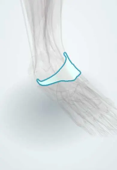
Fibular retinacula !
The fibular retinacula are bands behind the outer ankle bone that keep the fibularis tendons in their groove.
Official name: Superior fibular retinaculum
Opens in the Durgical app, no sign-up needed.
What it is
Two bands behind and below the outer ankle bone.
What it does
Keep the fibularis longus and brevis tendons in their groove behind the outer ankle bone.
Key facts
- Attaches
- Superior: lateral malleolus to calcaneus; inferior: lateral calcaneus (fibular trochlea), continuous with inferior extensor retinaculum
- Function
- Hold fibularis longus and brevis tendons behind lateral malleolus; superior tear lets them sublux
Relations
- Contents
- Superior retinaculum: fibularis brevis tendon (closer to the bone) and fibularis longus tendon (behind and outside it) in a common sheath; inferior retinaculum: the two tendons in separate sheaths
- Deep
- Retromalleolar groove of the fibula, deepened by a fibrocartilage ridge, then the CFL and the lateral wall of the calcaneus
- Behind
- Sural nerve and small saphenous vein running behind and below the lateral malleolus
- In front
- The inferior retinaculum continues into the stem of the inferior extensor retinaculum
How to find it
Behind and below the lateral malleolus. When the foot everts and dorsiflexes against resistance, the tendons stand out in their groove; in instability they can be seen or felt snapping forward over the malleolus.
Examination
- Resisted eversion and dorsiflexion from a plantarflexed, inverted start, with a finger on the groove: tendons slipping over the malleolus mean retinaculum failure
- Pressure on the groove during resisted eversion causing pain suggests a fibular tendon tear or tenosynovitis
On imaging
AP or mortise X-ray shows the fleck sign; dynamic ultrasound shows the tendons subluxing in real time; MRI shows tendon splits, a shallow or convex groove, a low-lying brevis muscle belly or an extra muscle.
Common problems
- Torn in some ankle injuries, letting the tendons slip out of the groove (subluxation), often with a snapping feeling. Surgery may be needed to repair it.
Clinical notes
- Acute fibular tendon dislocation is often diagnosed as a lateral ankle sprain at first
- Eckert–Davis grades run from the retinaculum stripping the fibular periosteum (I) to a cortical bone avulsion (III)
- Calcaneal fractures with lateral wall blow-out can dislocate the fibular tendons and tear the retinacula
- Repair reattaches the retinaculum and may deepen the groove
Anatomical variants
- An accessory fibularis quartus muscle or a low-lying fibularis brevis belly can crowd the groove; the groove itself may be flat or convex instead of concave
Test yourself
Think of the answer, then tap the question to see it.
What do the fibular retinacula hold, and which one is the main restraint?
The fibularis longus and brevis tendons behind the lateral malleolus; the superior fibular retinaculum is the main restraint.
What mechanism tears the superior fibular retinaculum?
Forced dorsiflexion and eversion while the fibularis muscles contract, as in skiing or football.
What does a fleck sign off the lateral rim of the fibula mean on an ankle X-ray?
Avulsion of the superior fibular retinaculum.
Under the inferior fibular retinaculum, what separates the fibularis brevis from the fibularis longus tendon?
The fibular trochlea of the calcaneus: brevis above, longus below.
Educational information, not medical advice. For any symptom, see a health professional.
Its names
- Official Latin (TA2)
- retinaculum fibulare superius; retinaculum fibulare inferius TA2 2715
- Official Spanish (DeCS)
- Retináculos fibulares superior e inferior
- English (TA2)
- Superior fibular retinaculum
- Also called
- superior peroneal retinaculum · inferior peroneal retinaculum · retinaculum proximale musculorum fibularium
Related
Sources: Terminologia Anatomica (TA2); Anatomy, Bony Pelvis and Lower Limb: Leg Lateral Compartment.
Spotted a mistake?We are always improving for the community. Tell us what is wrong and our team will review it.Report a mistake
Representative model, for educational purposes only.