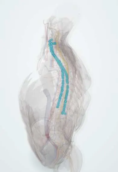
Sympathetic trunk !
The sympathetic trunk is a chain of nerve knots down each side of the front of the spine; it controls automatic functions such as sweating.
Opens in the Durgical app, no sign-up needed.
What it is
A chain of nerve knots running down each side of the front of the spine.
What it does
Controls automatic functions such as sweating, blood vessel tone and pupil size. Injury in the neck can cause a droopy eyelid (Horner syndrome); in the lower back, one leg may feel warmer.
Key facts
- Roots
- Preganglionic fibres from lateral horn T1–L2 via white rami; grey rami to every spinal nerve; skull base to coccyx (ganglion impar)
- Moves
- Sweat glands, vascular smooth muscle, arrector pili; cardiac branches; splanchnic nerves (T5–L2) to abdominal viscera; dilator pupillae (cervical ganglia)
- Feeling from
- Visceral pain afferents run with sympathetic fibres back to T1–L2 segments (referred pain)
Relations
- Course
- Neck: three ganglia (superior, middle, inferior or stellate) on the longus muscles; chest: over the rib heads; abdomen: on the vertebral bodies along the medial edge of psoas; pelvis: medial to the anterior sacral foramina, the two trunks joining at the ganglion impar in front of the coccyx
- Above
- Superior cervical ganglion (fused C1–C4, the largest, about 5 cm) at C2–C3, behind the internal carotid artery on longus capitis; the inconstant middle ganglion lies at C6 by the inferior thyroid artery; the ansa subclavia loops around the subclavian artery
- In front
- Neck: the carotid sheath; chest: the parietal pleura; abdomen: the inferior vena cava on the right, the aorta and para-aortic nodes on the left
- Behind
- Longus colli and longus capitis in the neck, the rib heads in the chest; the lumbar vessels usually pass behind it
- Lateral
- The psoas major in the abdomen; the posterior intercostal vessels and nerves in the chest
How to find it
In thoracoscopic surgery the thoracic trunk is seen through the pleura as a white cord over the rib heads, the second rib head marking T2. In the neck, the stellate ganglion block is guided to the anterior tubercle of C6 (Chassaignac tubercle), beside the trachea at the cricoid level.
Examination
- Pupils in dim light: the Horner pupil dilates slowly (dilation lag); eye drop tests confirm the diagnosis and help locate the lesion
- Sweat testing maps the area of anhidrosis, which is larger with lesions before the superior cervical ganglion
On imaging
Not seen directly; on CT it runs beside the vertebral bodies near the rib heads. Its ganglion tumours (neuroblastoma in young children, ganglioneuroma) are elongated paravertebral masses in the posterior mediastinum or retroperitoneum. In Horner syndrome imaging follows the whole path: brain and neck MRI, CT or MR angiography for carotid dissection, and the lung apex for a Pancoast tumour.
Clinical notes
- Causes along the path: lateral medullary stroke, cervical cord lesion, Pancoast tumour, carotid dissection (a painful Horner) and neck surgery or trauma; a new painful Horner prompts urgent carotid imaging
- Stellate ganglion block is used for complex regional pain syndrome of the arm and some refractory arrhythmias; risks include injection into the vertebral artery and hoarseness from recurrent laryngeal nerve block
- Thoracoscopic sympathectomy (T2–T3) treats severe palmar sweating; compensatory sweating elsewhere on the trunk is the main drawback
- Lumbar sympathectomy or block (about L2–L4) warms and dries the foot; removing the upper lumbar ganglia on both sides can impair ejaculation
Anatomical variants
- A fused stellate ganglion is present in about 85% of people (otherwise separate inferior cervical and T1 ganglia); a middle cervical ganglion in about 80%
- The number of lumbar ganglia on each side varies from 2 to 6, about 4 on average
Development
Neural crest cells form the chain ganglia and the adrenal medulla, which is why neuroblastoma can arise anywhere along the trunk or in the adrenal gland.
Related conditions
- Postural orthostatic tachycardia syndrome (POTS) and inappropriate sinus tachycardia
- Autonomic dysreflexia
- Horner syndrome
- Phaeochromocytoma
- Spinal cord injury
- Lung cancer
Test yourself
Think of the answer, then tap the question to see it.
Where do the preganglionic fibres of the sympathetic trunk arise?
In the lateral horn of the spinal cord at T1–L2, reaching the trunk through white rami; grey rami go back to every spinal nerve.
What are the features of Horner syndrome?
Ptosis, miosis and loss of facial sweating on one side, from a break anywhere in the three-neuron sympathetic path to the eye.
Where is the stellate ganglion, and what confirms a successful block?
In front of the C7 transverse process and neck of the first rib, behind the vertebral artery; a temporary Horner syndrome confirms the block.
What does a new painful Horner syndrome prompt?
Urgent carotid imaging, because carotid dissection is a cause of a painful Horner syndrome.
Educational information, not medical advice. For any symptom, see a health professional.
Its names
- Official Latin (TA2)
- truncus sympathicus; ganglia trunci sympathici TA2 6602
- Official Spanish (DeCS)
- Tronco simpático DeCS D013564
- English (TA2)
- Sympathetic trunk
- Also called
- sympathetic chain
Related
Sources: Terminologia Anatomica (TA2); DeCS; Anatomy, Head and Neck, Sympathetic Chain; Anatomy, Back, Lumbar Sympathetic Chain; Anatomía humana (4.ª ed.), tomo 1, cap. 41: Porción simpática; Tratado de anatomía humana (9.ª ed.), tomo II, Libro IV Angiología, sección 1, cap. I Corazón (arts. VI–IX) y cap. II Pericardio.
Spotted a mistake?We are always improving for the community. Tell us what is wrong and our team will review it.Report a mistake
Representative model, for educational purposes only.