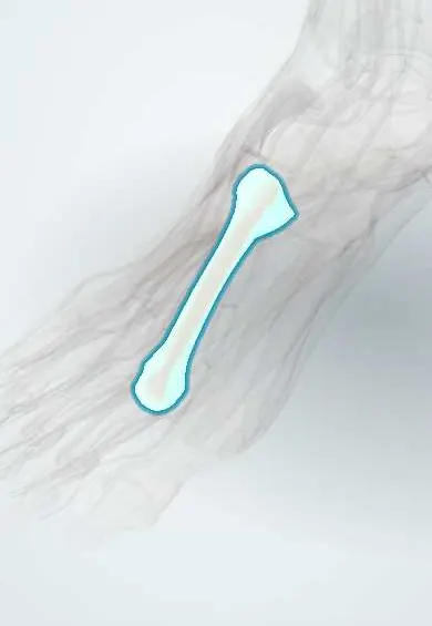
Metatarsal fractures (including stress fractures) !
Metatarsal fractures are breaks in the long bones of the forefoot, from an injury or from repeated loading (a stress or «march» fracture).
Updated 2026-10-29
Structures involved
Tap one to see what it is and open it in 3D.
- Second metatarsal
- Fifth metatarsal
- Third metatarsal
- Fibularis brevis
- First metatarsal
- Fourth metatarsal
- Lisfranc ligaments
- Plantar fascia
- Cuboid
- Dorsalis pedis artery
Why it happens
What it is
A break in one of the five metatarsal bones of the forefoot, either acute from injury or a stress fracture from repeated loading. The second metatarsal «march fracture» and fractures of the base of the fifth metatarsal are the classic types.
Who gets it
Metatarsal fractures are among the most common foot fractures. Stress fractures occur in runners, soldiers in training and dancers; acute fifth metatarsal fractures often follow an ankle twist.
Causes
- Inversion injury of the foot (avulsion of the fifth metatarsal base)
- Direct blow or crush injury
- Repeated loading with a sudden increase in training, such as marching or running (stress fracture)
- Low bone density (insufficiency fracture)
Risk factors
- Sudden increase in training volume or intensity
- Military training
- Low energy availability, menstrual disturbance or low bone density in athletes
- Low vitamin D
- Cavus or flat foot and hallux valgus that shift load
- Osteoporosis, diabetes with neuropathy
Pathophysiology
In a stress fracture, repeated loading causes microdamage faster than bone can remodel it. The long, fixed second metatarsal takes high loads at push-off, so it is the usual site. At the base of the fifth metatarsal, the pull of fibularis brevis and the plantar fascia causes avulsion, while the metaphysis–diaphysis junction (Jones zone) has a poor blood supply and heals slowly.
Classification
- Acute traumatic or stress fracture
- By bone (first to fifth) and site: head, neck, shaft or base
- Fifth metatarsal base: zone 1 tuberosity avulsion, zone 2 Jones fracture, zone 3 proximal shaft stress fracture
- Displaced or non-displaced; open or closed
Symptoms and signs
- Forefoot pain on weight bearing
- Local swelling and bruising after injury
- Point tenderness over the bone
- In stress fractures, pain that starts gradually with activity and later persists at rest
Diagnosis
- Clinical
- Point tenderness over the metatarsal; the Ottawa foot rules (tenderness at the fifth metatarsal base or navicular, or inability to bear weight) guide the need for X-rays.
- Tests
- In stress fractures, vitamin D and other bone health tests when risk factors are present.
- Imaging
- Foot X-rays in three views. Early stress fractures are often invisible on X-ray; MRI (or bone scan) shows them, and callus appears on X-ray weeks later. CT for complex patterns.
Differential diagnosis
- Lateral ankle sprain
- Lisfranc (tarsometatarsal) injury
- Morton neuroma
- Metatarsophalangeal synovitis or plantar plate injury
- Freiberg disease
- Tendinopathy of fibularis brevis
Goal of treatment
Achieve bone healing in good alignment, control pain and return safely to walking and sport while correcting the cause of overload.
Treatment
- Conservative
- Most non-displaced shaft fractures and fifth-metatarsal tuberosity avulsions: walking in a stiff-soled shoe or boot as pain allows.
- Conservative
- Jones fracture: a period of non-weight-bearing in a cast or boot because of the risk of delayed healing.
- Medical
- Simple analgesics; sources differ on NSAIDs during fracture healing. Treatment of low vitamin D or low bone density when present.
- Surgical
- Internal fixation for displaced or multiple fractures, and intramedullary screw fixation of Jones fractures in athletes or after nonunion.
- Rehabilitation
- Graded return to impact activity, review of training load, footwear and nutrition.
Complications
- Delayed union or nonunion, especially of the Jones zone
- Malunion with transfer pain under other metatarsal heads
- Refracture
- Compartment syndrome of the foot after crush injury
- Missed Lisfranc injury
Prognosis
Most metatarsal fractures heal well with protected walking. Jones fractures and stress fractures with ongoing risk factors are prone to slow healing and recurrence.
Prevention
- Gradual increase in training load
- Adequate energy intake, calcium and vitamin D
- Suitable footwear and rest days
Test yourself
Think of the answer, then tap the question to see it.
Which metatarsal is most often affected by a «march fracture»?
The second metatarsal, usually the shaft or neck, in recruits or runners after a rise in load.
What separates a Jones fracture from a tuberosity avulsion of the fifth metatarsal?
An avulsion is at the tuberosity tip (zone 1); a Jones fracture is at the metaphysis–diaphysis junction (zone 2), where poor blood supply slows healing.
Which tendon is classically blamed for avulsion of the fifth metatarsal base?
The fibularis (peroneus) brevis, with the lateral band of the plantar fascia, during an inversion injury.
Why may the first X-ray be normal in a metatarsal stress fracture?
Early stress fractures show no line; MRI detects them, and callus appears on X-ray only after some weeks.
Which clinical rule helps decide if foot X-rays are needed after injury?
The Ottawa foot rules: tenderness at the fifth metatarsal base or navicular, or inability to bear weight.
Related conditions
- Medial tibial stress syndrome (shin splints)
- Lateral Ankle Sprain
- Morton neuroma
- Acute compartment syndrome
- Osteoporosis
- Hallux valgus (bunion)
Educational information, not medical advice. For any symptom, see a health professional.
Sources: Metatarsal Fractures (StatPearls (NCBI Bookshelf), 2023).
Spotted a mistake?We are always improving for the community. Tell us what is wrong and our team will review it.Report a mistake
Representative model, for educational purposes only.