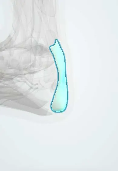
Flexor retinaculum !
The flexor retinaculum is a band behind the inner ankle bone that roofs the tarsal tunnel, where tendons, vessels and a nerve pass.
Official name: Flexor retinaculum of ankle
Opens in the Durgical app, no sign-up needed.
What it is
A band from the inner ankle bone to the heel bone. It forms the roof of the tarsal tunnel.
What it does
Holds in place the tendons, artery, veins and tibial nerve that pass behind the inner ankle.
Key facts
- Attaches
- Medial malleolus to medial process of calcaneus
- Function
- Roofs tarsal tunnel: tibialis posterior, FDL, posterior tibial vessels, tibial nerve, FHL (Tom, Dick And Very Nervous Harry)
Relations
- Contents
- Front to back: tibialis posterior tendon, flexor digitorum longus tendon, posterior tibial artery with its veins, tibial nerve, flexor hallucis longus tendon
- Deep
- Medial surface of the talus, sustentaculum tali and medial wall of the calcaneus (the floor of the tunnel)
- Superficial
- Skin and the medial calcaneal nerve branches, which may pierce it on their way to the heel
- Below
- Abductor hallucis, beneath which the medial and lateral plantar nerves and vessels enter the sole
How to find it
It spans the hollow between the medial malleolus and the heel. The posterior tibial artery pulse is felt under it, about halfway between the medial malleolus and the Achilles tendon.
Examination
- Tinel sign over the tarsal tunnel: tingling into the sole suggests tibial nerve irritation
- Dorsiflexion–eversion test: holding the ankle dorsiflexed and everted with the toes extended reproduces symptoms in tarsal tunnel syndrome
- Reduced sensation in the sole with a spared heel fits compression of the nerve within the tunnel
On imaging
Ultrasound or MRI looks for something occupying the tunnel: a ganglion, varicose veins, tenosynovitis or an accessory muscle. Nerve conduction studies support the diagnosis.
Common problems
- Tarsal tunnel syndrome: pressure on the nerve under this band causes burning or numbness in the sole.
Clinical notes
- Tarsal tunnel release divides the retinaculum and the fascia of the abductor hallucis to free the nerve and its branches
- The posterior tibial pulse and the tibial nerve block for foot surgery are found at this level
Anatomical variants
- Accessory muscles in the tunnel, such as the flexor digitorum accessorius longus, can compress the tibial nerve
Related conditions
Test yourself
Think of the answer, then tap the question to see it.
What are the attachments of the ankle flexor retinaculum, and what does it roof?
From the medial malleolus to the medial process of the calcaneus; it is the roof of the tarsal tunnel.
What passes under the flexor retinaculum, from front to back?
Tibialis posterior, flexor digitorum longus, posterior tibial artery and veins, tibial nerve, flexor hallucis longus («Tom, Dick And Very Nervous Harry»).
Why can heel sensation be spared in tarsal tunnel syndrome?
The medial calcaneal branch of the tibial nerve often leaves above the flexor retinaculum.
Which provocative test reproduces symptoms of tarsal tunnel syndrome besides the Tinel sign?
The dorsiflexion–eversion test: holding the ankle dorsiflexed and everted with the toes extended.
Educational information, not medical advice. For any symptom, see a health professional.
Its names
- Official Latin (TA2)
- retinaculum flexorium tali TA2 2714
- Official Spanish (DeCS)
- Retináculo flexor del tobillo
- English (TA2)
- Flexor retinaculum of ankle
- Also called
- laciniate ligament · ligamentum laciniatum
Related
Sources: Terminologia Anatomica (TA2); Anatomy, Bony Pelvis and Lower Limb: Leg Posterior Compartment.
Spotted a mistake?We are always improving for the community. Tell us what is wrong and our team will review it.Report a mistake
Representative model, for educational purposes only.