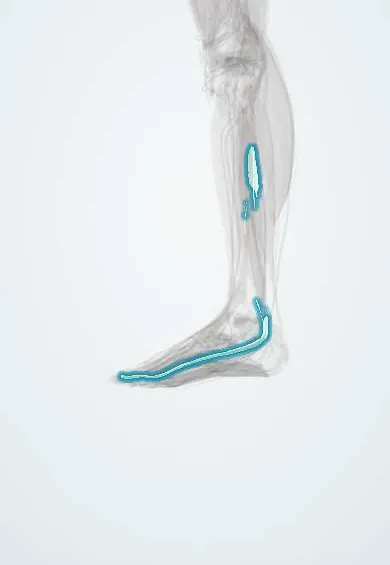
Big toe flexor tendon !
The big toe flexor tendon runs behind the ankle and under the sole to the tip of the big toe, which it curls; it helps push off.
Official name: Tendon of flexor hallucis longus
Opens in the Durgical app, no sign-up needed.
What it is
The tendon of the long big-toe flexor. It runs in a groove behind the talus and under the sole to the tip of the big toe.
What it does
Curls the big toe and helps push off. It is sometimes moved to the heel to reinforce a damaged Achilles tendon.
Key facts
- Attaches
- In a groove on the back of the talus between its two tubercles, under the sustentaculum tali, crossed by the FDL in the sole, then between the two hallux sesamoids to the base of the distal phalanx of the big toe
- Function
- Flexes the big toe for push-off; supports the medial arch; tendinopathy in dancers; used as a transfer to the calcaneus in chronic Achilles rupture
- Tight in
- Big toe extension with ankle dorsiflexion
Relations
- Course
- From the lower fibula it passes behind the lower tibia into the groove between the talar tubercles, under the sustentaculum tali, across the sole and on to the big toe
- Medial
- Posterior tibial artery and veins and the tibial nerve; further medial, the flexor digitorum longus and tibialis posterior tendons
- Lateral
- Lateral talar tubercle and, when present, an os trigonum
- Above
- Sustentaculum tali of the calcaneus, which acts as a pulley above it
How to find it
Bending the big toe against resistance makes it move deep behind the medial ankle, behind the neurovascular bundle; it is easier to feel under the first metatarsal head, between the sesamoids.
Examination
- Big-toe extension tested with the ankle pointed down and then pulled up: less extension with the ankle up suggests the tendon is tethered or narrowed behind the ankle
On imaging
Its sheath often connects with the ankle joint, so a little fluid around it is normal; disproportionate fluid, thickening or a split on MRI or ultrasound points to tenosynovitis; a lateral radiograph shows an os trigonum.
Common problems
- Tendinitis behind the inner ankle, common in ballet dancers ('dancer's tendinitis').
Clinical notes
- Transferred to the calcaneus to reconstruct chronic Achilles tendon ruptures and severe insertional Achilles disease: it is strong, works in phase with the calf and lies close by
- Releasing its tunnel behind the talus, often with removal of an os trigonum, treats stenosing tenosynovitis in dancers
- The posterolateral approach to posterior malleolus fractures uses the interval between the fibular tendons and the flexor hallucis longus, whose belly is lifted off the back of the tibia
Anatomical variants
- A slip joining it to the flexor digitorum longus in the sole is common
- A rare accessory muscle (peroneocalcaneus internus) can run beside it behind the ankle and be mistaken for it
Memory aid
'Harry' comes last in 'Tom, Dick And Very Nervous Harry': the flexor hallucis longus is the most posterior structure behind the medial malleolus.
Test yourself
Think of the answer, then tap the question to see it.
Which tendon is the most posterior structure behind the medial malleolus?
The flexor hallucis longus ('Harry' in 'Tom, Dick And Very Nervous Harry').
Why does posterior ankle arthroscopy work stay lateral to the flexor hallucis longus?
The tibial nerve and posterior tibial artery lie just medial to it.
What is hallux saltans in ballet dancers?
The thickened flexor hallucis longus catches in its tunnel behind the talus, making the big toe lock and then release.
Which tendon is transferred to the calcaneus in chronic Achilles tendon ruptures?
The flexor hallucis longus: it is strong, works in phase with the calf and lies close by.
Educational information, not medical advice. For any symptom, see a health professional.
Its names
- Official Latin (TA2)
- flexor longus hallucis TA2 2668
- Official Spanish (DeCS)
- Tendón del flexor largo del dedo gordo DeCS D013710
- English (TA2)
- Tendon of flexor hallucis longus
- Also called
- flexor hallucis longus muscle
Related
Sources: Terminologia Anatomica (TA2); DeCS; Anatomy, Bony Pelvis and Lower Limb: Calf Flexor Hallucis Longus Muscle.
Spotted a mistake?We are always improving for the community. Tell us what is wrong and our team will review it.Report a mistake
Representative model, for educational purposes only.