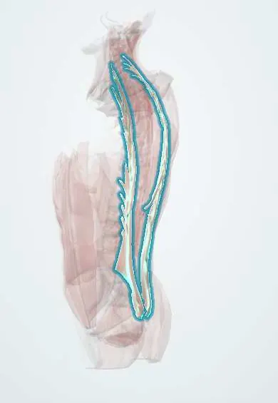
Iliocostalis !
The iliocostalis is the outer column of the erector spinae, from the pelvis to the ribs and neck; it straightens and holds the spine upright.
Official name: Iliocostalis muscles
Opens in the Durgical app, no sign-up needed.
What it is
The outer column of the erector spinae, running from the pelvis to the ribs and up to the neck.
What it does
Straightens the spine and holds it upright; on one side it bends the trunk to that side.
Key facts
- Origin
- Common erector spinae tendon: posterior iliac crest, sacrum, lumbar spinous processes; ribs (thoracis, cervicis parts)
- Insertion
- Angles of the ribs; transverse processes of C4–C6 (cervicis; some texts: C4–C7)
- Action
- Bilateral: extends the vertebral column; unilateral: lateral flexion to the same side
- Nerve
- Dorsal rami of spinal nerves (lateral branches; lower cervical, thoracic and lumbar)
- Blood supply
- Dorsal branches of posterior intercostal, subcostal and lumbar arteries
Relations
- Superficial
- Thoracolumbar fascia, latissimus dorsi and serratus posterior inferior; higher up, the rhomboids and trapezius
- Deep
- Ribs near their angles, with the levatores costarum and external intercostals; in the loin, the middle layer of the thoracolumbar fascia and quadratus lumborum
- Medial
- Longissimus, from which it is separated by a fat-filled cleft carrying lateral branches of the dorsal rami
How to find it
Lying face down and lifting the chest slightly, the erector spinae forms a ridge on each side of the spine; the iliocostalis is its outer part, felt over the rib angles in the thoracic region and at the lateral edge of the ridge in the loin.
On imaging
On axial MRI or CT it is the lateral column of the erector spinae, lateral to longissimus, reaching the rib angles in the thorax. It is rarely imaged on its own: ultrasound shows it as the outer part of the paraspinal mass during back blocks, and MRI shows a strain as feathery oedema or a haematoma after injury. In chronic back pain, MRI or CT can show fatty replacement of the paraspinal muscles.
Common problems
- Muscle strain and spasm: one of the most common causes of sudden low back pain.
- Weakness and fatigue add to chronic back pain.
How it is diagnosed
History and exam; imaging is not needed for a simple strain.
Treatment
- Staying active, heat, pain medicine and an exercise programme. Most strains settle in days to weeks.
Clinical notes
- Erector spinae plane block: under ultrasound, local anaesthetic placed deep to the erector spinae on a transverse process spreads over several levels to reach the spinal nerves' rami
- An intercostal nerve block is usually given at the angle of the rib, just lateral to the edge of iliocostalis, where the nerve still lies in the costal groove
- Fatty change and thinning of the paraspinal muscles on MRI are linked to chronic low back pain and to the loss of lumbar lordosis in adult spinal deformity
Development
A true (epaxial) back muscle: it forms from the dorsal part of the somite myotomes, which is why it is supplied by dorsal rami. Its long column arises when several neighbouring myotomes fuse and then split lengthwise into the three erector spinae columns.
Memory aid
I Love Spaghetti, from lateral to medial: Iliocostalis, Longissimus, Spinalis.
Related conditions
Test yourself
Think of the answer, then tap the question to see it.
What is the order of the erector spinae columns from lateral to medial?
Iliocostalis, longissimus, spinalis («I Love Spaghetti»); the iliocostalis is the lateral column.
What are the action and nerve supply of the iliocostalis?
Bilaterally it extends the spine; unilaterally it flexes it laterally to the same side. Supplied by lateral branches of the dorsal rami.
What is flexion-relaxation in the iliocostalis and the erector spinae?
The erector spinae go electrically quiet near full forward flexion, when ligaments and discs take the load.
Educational information, not medical advice. For any symptom, see a health professional.
Its names
- Official Latin (TA2)
- musculi iliocostales TA2 2257
- Official Spanish (DeCS)
- Músculo iliocostal DeCS D064170
- English (TA2)
- Iliocostalis muscles
- Also called
- iliocostalis muscle
Related
Sources: Terminologia Anatomica (TA2); DeCS; Anatomy, Back; Anatomy of the Human Body (20th ed.): Development of the Muscles (Lea & Febiger (Gray's Anatomy, 1918), 1918) · adapted, Public domain.
Spotted a mistake?We are always improving for the community. Tell us what is wrong and our team will review it.Report a mistake
Representative model, for educational purposes only.