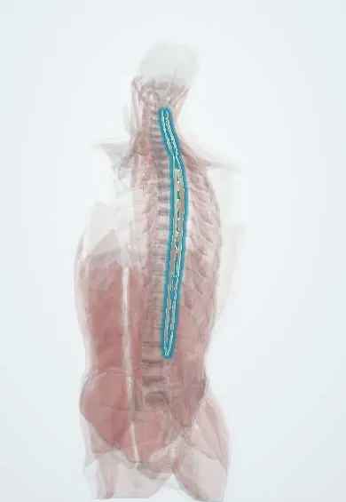
Spinalis !
The spinalis is the thin inner column of the erector spinae, running between the spinous processes; it helps straighten the spine.
Official name: Spinalis muscles
Opens in the Durgical app, no sign-up needed.
What it is
The thin inner column of the erector spinae, running between the spinous processes.
What it does
Helps straighten the spine.
Key facts
- Origin
- Spinous processes of T11–L2 (thoracis); nuchal ligament and C7 spine (cervicis, often absent)
- Insertion
- Spinous processes of upper thoracic vertebrae (about T2–T8); C2 spinous process (cervicis)
- Action
- Bilateral: extends the vertebral column; unilateral: lateral flexion
- Nerve
- Dorsal rami of spinal nerves (segmental; cervical and thoracic)
- Blood supply
- Dorsal branches of posterior intercostal and lumbar arteries
Relations
- Lateral
- Longissimus
- Medial
- Thoracic spinous processes and the supraspinous ligament
- Superficial
- Thoracolumbar fascia, trapezius, the rhomboids and latissimus dorsi
- Deep
- Semispinalis and multifidus (the transversospinal group) on the laminae
How to find it
It cannot be told apart from the rest of the erector spinae by touch. In a posterior midline approach to the thoracic spine it is the first muscle lifted off the sides of the spinous processes, with the transversospinal muscles beneath.
On imaging
Too thin to separate reliably on imaging; on axial MRI or CT of the thoracic spine it is the small medial part of the erector spinae against the sides of the spinous processes. After midline posterior surgery, MRI or CT may show scarring, atrophy or fatty change in the paraspinal muscles at the operated levels. Back pain imaging looks at the spine and whole erector spinae.
Common problems
- Muscle strain and spasm: one of the most common causes of sudden low back pain.
- Weakness and fatigue add to chronic back pain.
How it is diagnosed
History and exam; imaging is not needed for a simple strain.
Treatment
- Staying active, heat, pain medicine and an exercise programme. Most strains settle in days to weeks.
Clinical notes
- Detached from the spinous processes in midline posterior thoracic approaches; scarring and loss of nerve supply to the paraspinal muscles are thought to add to back pain after open surgery
- Percutaneous pedicle screws and tubular retractors pass through the paraspinal muscles instead of lifting them off the spinous processes, which lessens damage to it and its neighbours
Development
Arises, like the rest of the erector spinae, from fused dorsal (epaxial) myotomes, and keeps their dorsal ramus nerve supply.
Memory aid
I Love Spaghetti, from lateral to medial: Iliocostalis, Longissimus, Spinalis.
Test yourself
Think of the answer, then tap the question to see it.
Where does the spinalis lie within the erector spinae?
It is the smallest and most medial column, running from spinous process to spinous process.
Which part of the spinalis is the only constant one?
Spinalis thoracis; spinalis cervicis is often absent and spinalis capitis usually blends with semispinalis capitis.
In which approach is the spinalis the first muscle lifted off the spinous processes?
The midline posterior approach to the thoracic spine; the transversospinal muscles lie beneath it.
Educational information, not medical advice. For any symptom, see a health professional.
Its names
- Official Latin (TA2)
- musculi spinales TA2 2267
- Official Spanish (DeCS)
- Músculo espinoso DeCS D064170
- English (TA2)
- Spinalis muscles
- Also called
- spinalis muscle · musculus spinalis dorsi
Related
Sources: Terminologia Anatomica (TA2); DeCS; Anatomy, Back; Anatomy of the Human Body (20th ed.): Development of the Muscles (Lea & Febiger (Gray's Anatomy, 1918), 1918) · adapted, Public domain.
Spotted a mistake?We are always improving for the community. Tell us what is wrong and our team will review it.Report a mistake
Representative model, for educational purposes only.