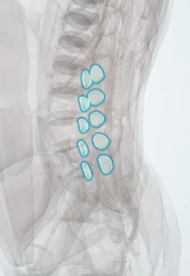
Mechanical low back pain !
Mechanical low back pain comes from the muscles, joints or discs of the lower back and changes with movement; usually no single cause is found.
Updated 2026-10-29
Structures involved
Tap one to see what it is and open it in 3D.
- Low-back facet joints
- L4–L5 disc
- L5–S1 disc
- Multifidus
- L4 vertebra
- L5 vertebra
- Iliocostalis
- Longissimus
- Quadratus lumborum
- Thoracolumbar fascia
In the clinical exam
Why it happens
What it is
Pain between the lower ribs and the gluteal folds that comes from the spine's muscles, ligaments, joints or discs and changes with posture and movement. Most cases are nonspecific: no single structural cause is found.
Who gets it
One of the most common reasons for primary care visits and work absence worldwide, including in Colombia and the rest of Latin America. Most adults have at least one episode in their lives.
Causes
- Nonspecific muscle and ligament strain, often after lifting, twisting or prolonged posture
- Degenerative changes of the discs and facet joints
- Sacroiliac joint pain
- Often no identifiable cause (nonspecific low back pain)
Risk factors
- Heavy lifting, bending and twisting at work
- Physical inactivity and poor trunk conditioning
- Obesity
- Smoking
- Previous episodes of low back pain
- Psychosocial factors: depression, job dissatisfaction, fear of movement
Pathophysiology
Pain arises from sensitised structures such as paraspinal muscles, the thoracolumbar fascia, facet joint capsules and the outer annulus of the disc, which carry nociceptive nerve endings. Muscle spasm and guarding add to it. In chronic pain, central sensitisation and psychosocial factors (yellow flags) often matter more than the tissue findings.
Classification
- Acute: under 6 weeks
- Subacute: 6 to 12 weeks
- Chronic: over 12 weeks
- Triage groups: nonspecific, with radiculopathy, or suspected serious pathology
Symptoms and signs
- Aching low back pain, sometimes spreading to the buttock or thigh but not below the knee
- Pain worse with movement, bending or sitting and eased by rest or position change
- Stiffness and paraspinal muscle spasm
- No neurological deficit in the legs
Diagnosis
- Clinical
- A careful history and examination, with screening for red flags and for yellow flags (fear-avoidance, low mood, beliefs that pain means harm). Neurological examination of the legs and straight leg raise to detect radiculopathy.
- Tests
- Usually none. Blood count, ESR or CRP when infection or malignancy is suspected.
- Imaging
- Not routine in acute nonspecific pain without red flags; it does not change outcomes and degenerative findings are common in people without pain. X-ray, CT or MRI when red flags are present or pain persists despite care (MRI is preferred for neural or soft tissue concerns).
Differential diagnosis
- Lumbar disc herniation with radiculopathy
- Lumbar spinal stenosis
- Vertebral compression fracture
- Spinal infection or metastasis
- Axial spondyloarthritis
- Referred pain: kidney stones, pyelonephritis, abdominal aortic aneurysm
Goal of treatment
Relieve pain, keep the person active and working, and prevent the change to chronic pain and disability, while not missing serious causes.
Treatment
- Conservative
- Reassurance and education about the usually good course, with advice to stay active and avoid bed rest. Superficial heat may help in acute pain.
- Medical
- NSAIDs for short periods, sometimes with a muscle relaxant. Opioids are used sparingly and briefly, if at all. Guidelines differ on the value of simple analgesics.
- Rehabilitation
- Exercise therapy, physical therapy and core strengthening, mainly for subacute and chronic pain; spinal manipulation is an option in some guidelines.
- Lifestyle
- Graded return to work, weight control, stopping smoking and regular activity. Psychological approaches such as cognitive behavioural therapy help in chronic pain.
Drugs for this condition
Complications
- Chronic low back pain
- Disability and long work absence
- Depression and anxiety
- Adverse effects of analgesics and opioid dependence
Prognosis
Most acute episodes improve within a few weeks, but recurrence is common. A minority develop chronic pain, and yellow flags predict this better than imaging findings.
Prevention
- Regular exercise and trunk strengthening
- Lifting technique and workplace ergonomics
- Healthy weight and not smoking
Test yourself
Think of the answer, then tap the question to see it.
Is imaging recommended in acute nonspecific low back pain without red flags?
No. Imaging is not routine: it does not improve outcomes and degenerative findings are common in people without pain.
Which red flags in low back pain suggest cauda equina syndrome?
Saddle anaesthesia, new urinary retention or incontinence, faecal incontinence and bilateral or progressive leg weakness.
What are yellow flags in low back pain?
Psychosocial factors such as fear-avoidance, catastrophising, low mood and work problems that predict chronic pain and disability.
What general advice about activity is given in acute mechanical low back pain?
To stay as active as tolerated and avoid bed rest, which delays recovery.
After how long is low back pain called chronic?
After more than 12 weeks (3 months).
Related conditions
- Lumbar Disc Herniation
- Sciatica (Lumbar Radiculopathy)
- Lumbar spinal stenosis
- Cauda equina syndrome
- Osteoporotic vertebral compression fracture
- Spondylolisthesis
- Scoliosis
- Abdominal aortic aneurysm
Educational information, not medical advice. For any symptom, see a health professional.
Sources: Low Back Pain: Evaluation and Management (StatPearls (NCBI Bookshelf), 2025).
Spotted a mistake?We are always improving for the community. Tell us what is wrong and our team will review it.Report a mistake
Representative model, for educational purposes only.