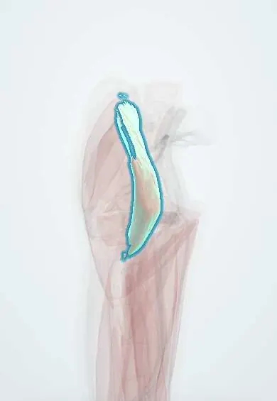
Iliacus !
The iliacus is a fan-shaped muscle lining the inside of the pelvis; together with the psoas it lifts the thigh.
Official name: Iliacus muscle
Opens in the Durgical app, no sign-up needed.
What it is
A fan-shaped muscle lining the inside of the pelvic wing (the iliac fossa). It joins the psoas in a shared tendon to the femur.
What it does
Lifts the thigh (bends the hip) together with the psoas.
Key facts
- Origin
- Iliac fossa; inner lip of iliac crest; ala of sacrum
- Insertion
- Lesser trochanter (via psoas tendon) and femur just below it
- Action
- Flexes hip; stabilizes hip joint
- Nerve
- Femoral nerve (L2–L3)
- Blood supply
- Iliolumbar artery (iliac branch); deep circumflex iliac artery; obturator artery
Relations
- In front
- Iliac fascia; the caecum on the right, the descending and sigmoid colon on the left; the lateral femoral cutaneous nerve crosses it toward the anterior superior iliac spine
- Medial
- Psoas major, with the femoral nerve in the groove between them
- Deep
- The iliac fossa of the hip bone
- Below
- Leaves the pelvis under the inguinal ligament, lateral to the psoas, then lies on the front of the hip capsule, separated from it by the iliopsoas bursa
How to find it
With the hip slightly flexed to relax the abdominal wall, deep pressure just inside the iliac crest and the anterior superior iliac spine reaches the muscle lining the iliac fossa. In the groin it lies lateral to the psoas, under the femoral nerve.
Examination
- Thomas test: lying supine, the opposite knee is held to the chest to flatten the lumbar curve; if the tested thigh lifts off the couch, there is a hip flexion contracture or a short iliopsoas
- FABER (Patrick) test: the hip is placed in flexion, abduction and external rotation and the knee lowered gently; groin pain points mainly to the hip joint, a differential of iliopsoas pain, and pain at the back of the pelvis to the sacroiliac joint
On imaging
CT is the first test for bleeding or abscess: it shows the muscle lining the iliac fossa lateral to the psoas. A haematoma, often on anticoagulants, enlarges it and can compress the femoral nerve; an abscess shows as fluid with an enhancing rim. MRI shows strains and tumours. Ultrasound is used for the fascia iliaca block. Radiographs do not show it.
Common problems
- Strain or tendinitis from kicking, sprinting or repeated hip bending.
- Snapping hip (inside type): the tendon catches over the front of the pelvis with a clunk.
- Tight hip flexors from long sitting can add to back or hip pain.
- Uncommonly, tendon irritation after hip replacement if it rubs on the implant.
How it is diagnosed
Physical exam (pain when lifting the leg against resistance); ultrasound, which can also show the snapping during movement; MRI.
Treatment
- Rest from the painful activity, stretching and progressive strengthening with a physical therapist.
- Ultrasound-guided injection into the iliopsoas bursa or around the tendon.
- Rarely, surgery to lengthen or release the tendon when snapping or pain does not improve.
Clinical notes
- Iliacus haematoma (anticoagulation, haemophilia, trauma): groin pain with the hip held flexed, weak knee extension and numbness of the front of the thigh from femoral nerve compression
- Anterior iliac crest bone graft is taken from the inner table after the iliacus is stripped; the lateral femoral cutaneous nerve near the anterior superior iliac spine is at risk
Anatomical variants
- Accessory slips of the iliacus, separate from the main belly, are described
Development
Belongs to the dorsal muscle mass of the limb bud, hence its femoral nerve supply from the posterior divisions of L2–L4. The 90° medial rotation of the lower limb in the seventh week brings these dorsal-mass muscles round to the front of the hip and thigh.
Related conditions
Test yourself
Think of the answer, then tap the question to see it.
Which nerve supplies the iliacus, and what are its actions?
The femoral nerve (L2–L3); the iliacus flexes the hip and stabilizes the hip joint.
Why can an iliacus haematoma cause knee extension weakness?
The femoral nerve runs in the groove between the iliacus and psoas under the iliac fascia, so bleeding compresses it.
What is the fascia iliaca block, and what is it used for?
Local anaesthetic under the fascia over the iliacus spreads to the femoral and lateral femoral cutaneous nerves; used for hip fracture pain.
Which nerve is at risk when the iliacus is stripped for an anterior iliac crest bone graft?
The lateral femoral cutaneous nerve, near the anterior superior iliac spine.
Educational information, not medical advice. For any symptom, see a health professional.
Its names
- Official Latin (TA2)
- musculus iliacus TA2 2594
- Official Spanish (DeCS)
- Músculo ilíaco
- English (TA2)
- Iliacus muscle
- Also called
- musculus ilicus
Related
Sources: Terminologia Anatomica (TA2); Anatomy, Bony Pelvis and Lower Limb: Iliopsoas Muscle; 8.5 Development of the Appendicular Skeleton (OpenStax (Rice University), 2022) · adapted, CC BY 4.0.
Spotted a mistake?We are always improving for the community. Tell us what is wrong and our team will review it.Report a mistake
Representative model, for educational purposes only.