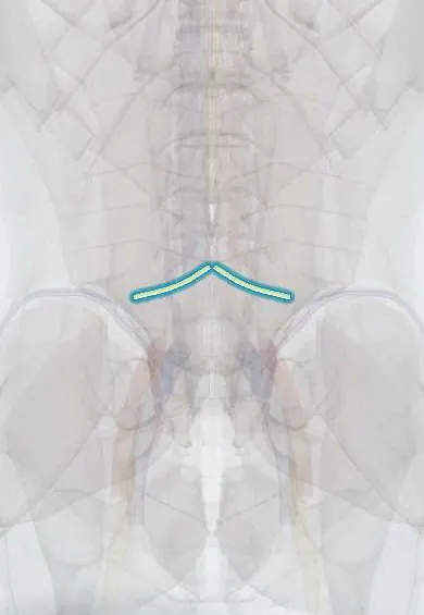
L4 nerve root !
The L4 nerve root leaves the spine through the L4-L5 opening; it helps straighten the knee and gives the knee-jerk reflex.
Official name: L4
Opens in the Durgical app, no sign-up needed.
What it is
The L4 nerve root, leaving the spine through the L4-L5 opening.
What it does
Helps straighten the knee (quadriceps), gives the knee-jerk reflex, and feeling over the inner shin.
Key facts
- Roots
- L4 dorsal and ventral roots; exits the L4–L5 foramen; compressed by L3–L4 posterolateral herniation (or far-lateral L4–L5)
- Moves
- Quadriceps (knee extension); tibialis anterior (with L5); hip adductors; patellar reflex (L3–L4)
- Feeling from
- Medial leg and medial malleolus
Relations
- Course
- Crosses behind the L3–L4 disc inside the dural sac (where a posterolateral herniation catches it), passes under the L4 pedicle and leaves the L4–L5 foramen to join the femoral and obturator nerves and the lumbosacral trunk
- In front
- The L4 vertebral body and, in the foramen, the L4–L5 disc
- Behind
- The L4–L5 facet joint and ligamentum flavum
- Lateral
- The psoas major, inside which the lumbar plexus forms
How to find it
It cannot be felt. It leaves through the L4–L5 foramen, under the L4 pedicle; in the paraspinal (Wiltse) approach for a far-lateral disc it is found lateral to the pars interarticularis, between the L4 and L5 transverse processes.
Examination
- Femoral nerve stretch test: lying prone, the knee is bent and the hip extended; pain in the front of the thigh suggests L2–L4 root tension
- Patellar reflex, with the Jendrassik manoeuvre (hooking the fingers and pulling) if it seems absent
- Heel walking and resisted ankle dorsiflexion (L4 key muscle); repeated single-leg sit-to-stand shows mild quadriceps weakness
- Pinprick at the medial malleolus (L4 key point)
On imaging
MRI is the test of choice. Axial T2 at L3–L4 shows a paracentral herniation pressing on the traversing L4 root in the lateral recess; at L4–L5 a foraminal or far-lateral herniation presses on it as the exiting root. Sagittal slices through the foramen show foraminal narrowing, often with disc loss or spondylolisthesis. Radiographs only show bony changes.
Common problems
- Pinched by an L3-L4 herniation or by a far-lateral L4-L5 herniation: pain in the front of the thigh and inner shin.
How it is diagnosed
Exam (straight-leg raise, big toe and foot lift strength, feeling); MRI.
Treatment
- Most sciatica improves within weeks to months with activity, physical therapy, medicine or an epidural injection.
- Severe, lasting pain or weakness: microdiscectomy.
Clinical notes
- L4 root vs femoral nerve: a femoral lesion spares the hip adductors (obturator nerve) and tibialis anterior and causes no back pain; an L4 root lesion can weaken both
- Far-lateral L4–L5 herniation catches the exiting L4 root and its ganglion outside the canal, with intense pain; it is easy to miss without looking lateral to the foramen on MRI
- Diabetic lumbosacral radiculoplexus neuropathy (diabetic amyotrophy) mimics an upper lumbar radiculopathy: severe thigh pain, wasting and weight loss
Related conditions
Test yourself
Think of the answer, then tap the question to see it.
Which disc herniations compress the L4 nerve root?
A posterolateral L3–L4 herniation, or a far-lateral L4–L5 herniation that catches the exiting L4 root outside the canal.
Which reflex is reduced with an L4 radiculopathy?
The patellar (knee-jerk) reflex (L3–L4), with pain from the front of the thigh to the inner shin.
Which tension test is used for the L2–L4 roots instead of the straight-leg raise?
The femoral nerve stretch test: prone, knee bent and hip extended; pain in the front of the thigh suggests L2–L4 root tension.
How is an L4 root lesion told apart from a femoral nerve lesion?
A femoral lesion spares the hip adductors (obturator nerve) and tibialis anterior and causes no back pain; an L4 root lesion can weaken both.
Educational information, not medical advice. For any symptom, see a health professional.
Its names
- Official Latin (TA2)
- radix nervi spinalis L4 TA2 6489
- Official Spanish (DeCS)
- Raíz L4 DeCS D013126
- English (TA2)
- L4
- Also called
- spinal nerves L1-L5
Related
Sources: Terminologia Anatomica (TA2); DeCS; Anatomy, Skin, Dermatomes; Neuroanatomy, Spinal Nerves.
Spotted a mistake?We are always improving for the community. Tell us what is wrong and our team will review it.Report a mistake
Representative model, for educational purposes only.