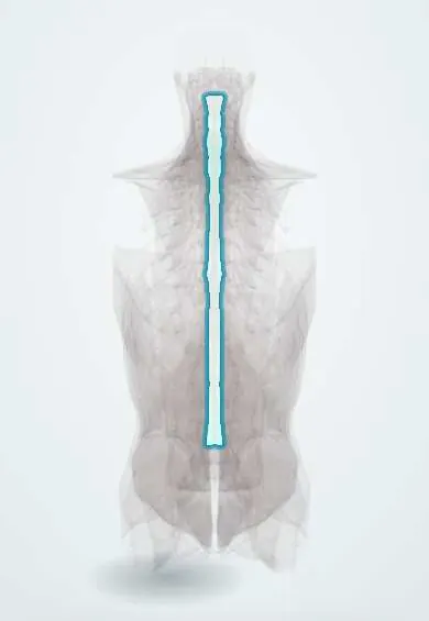
Ligamentum flavum !
The ligamentum flavum is a set of short, yellow, elastic ligaments joining the back arches of the vertebrae, forming the back wall of the canal.
Official name: Ligamenta flava
Opens in the Durgical app, no sign-up needed.
What it is
Short, yellow, elastic ligaments joining the laminae (back arches) of neighbouring vertebrae, forming the back wall of the spinal canal.
What it does
Stretches when you bend forward and springs back as you straighten, keeping the canal wall smooth.
Key facts
- Attaches
- Laminae of adjacent vertebrae (front of lamina above to back of upper edge of lamina below), C2–S1; forms back wall of canal
- Function
- Elastic (yellow elastin); limits abrupt flexion and helps return to upright; buckling or thickening narrows the canal (stenosis); pierced in lumbar puncture
- Tight in
- Flexion
Relations
- Course
- Midline lumbar puncture needle path: skin, subcutaneous fat, supraspinous ligament, interspinous ligament, ligamentum flavum (the 'give'), epidural space, dura and arachnoid, then CSF in the subarachnoid space
- In front
- Posterior epidural space with fat and veins, then the dural sac
- Behind
- Laminae and, in the midline, the interspinous ligament
- Lateral
- Front of the facet joint capsules, into which it blends, and the back edge of the intervertebral foramen
How to find it
From behind, the laminae overlap most of it, so only a short strip shows in each gap between laminae, widest in the lumbar spine with the trunk bent forward. Its full height is seen from inside the canal in dissection, running from the front of the lamina above to the upper edge of the lamina below.
On imaging
Axial T2 MRI shows it as a dark V lining the back of the canal; thickening and buckling with facet hypertrophy narrow the canal into a trefoil shape. CT shows calcification and ossification of the ligamentum flavum, which occurs mostly in the lower thoracic spine.
Common problems
- Thickens and buckles with age, narrowing the canal: a main cause of lumbar spinal stenosis.
- Leg pain and heaviness when standing or walking that eases when sitting or leaning forward (neurogenic claudication).
How it is diagnosed
MRI shows the thickened ligament and how narrow the canal is.
Treatment
- Physical therapy, walking programmes, medicine and injections.
- Surgery (laminectomy) removes the thickened ligament and part of the bone to open the canal.
Clinical notes
- Neurogenic claudication: standing and walking (extension) buckle it into the canal; sitting or leaning forward stretches it and opens the canal (the shopping-cart sign)
- Ossification of the ligamentum flavum, more common in East Asian populations and mainly in the lower thoracic spine, causes thoracic myelopathy, sometimes together with OPLL
- Laminectomy and interlaminar approaches remove part of it (flavectomy) to decompress the canal or reach a disc
Anatomical variants
- Midline gaps between the left and right halves are common, especially in the cervical and upper thoracic spine, so the loss of resistance may be less clear there
Memory aid
Flavum = yellow = elastic: the yellow ligament snaps back like a rubber band.
Related conditions
Test yourself
Think of the answer, then tap the question to see it.
Where does the ligamentum flavum attach?
Between the laminae of adjacent vertebrae (front of the lamina above to the back of the upper edge of the lamina below), from C2 to S1, forming the back wall of the canal.
Why is the ligamentum flavum yellow and elastic, and what does that do?
It is very rich in elastin (about 80%), which keeps it taut even at rest so it does not buckle into the canal and helps the spine return upright from flexion.
What does the needle feel when it passes the ligamentum flavum during lumbar puncture or epidural placement?
Firm resistance that suddenly gives way as the needle enters the epidural space (loss of resistance).
How does the ligamentum flavum explain neurogenic claudication?
Standing and walking (extension) buckle the thickened ligament into the canal; sitting or leaning forward stretches it and opens the canal.
Where and in whom does ossification of the ligamentum flavum occur, and what does it cause?
Mainly in the lower thoracic spine, more often in East Asian populations; it causes thoracic myelopathy, sometimes with OPLL.
Educational information, not medical advice. For any symptom, see a health professional.
Its names
- Official Latin (TA2)
- ligamenta flava TA2 1675
- Official Spanish (DeCS)
- Ligamento amarillo DeCS D017843
- English (TA2)
- Ligamenta flava
- Also called
- ligamenta interarcualia
Related
- Dura (dural sac)
- Low-back facet joints
- Atlas (C1)
- Axis (C2)
- C3 vertebra
- C4 vertebra
- C5 vertebra
- C6 vertebra
Sources: Terminologia Anatomica (TA2); DeCS; Anatomy, Back, Lumbar Spine; Anatomy, Back, Posterior Longitudinal Ligament; Anatomy of the Human Body (20th ed.): Articulations of the Vertebral Column (Lea & Febiger (Gray's Anatomy, 1918), 1918) · adapted, Public domain.
Spotted a mistake?We are always improving for the community. Tell us what is wrong and our team will review it.Report a mistake
Representative model, for educational purposes only.