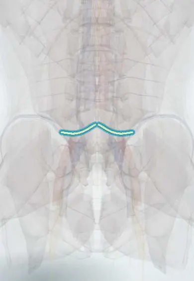
L5 nerve root !
The L5 nerve root passes behind the L4-L5 disc and leaves at L5-S1; it lifts the foot and big toe and gives feeling to the top of the foot.
Official name: L5
Opens in the Durgical app, no sign-up needed.
What it is
The L5 nerve root: it runs down past the back of the L4-L5 disc and leaves the spine through the L5-S1 opening.
What it does
Lifts the foot and big toe, and gives feeling over the outer leg and the top of the foot.
Key facts
- Roots
- L5 dorsal and ventral roots; exits the L5–S1 foramen; compressed by L4–L5 posterolateral herniation
- Moves
- Extensor hallucis longus, tibialis anterior (with L4), gluteus medius (hip abduction); no reliable reflex (some texts: medial hamstring)
- Feeling from
- Lateral leg, dorsum of foot and big toe (first web space)
Relations
- Course
- Passes behind the L4–L5 disc in the lateral recess, under the L5 pedicle and out of the L5–S1 foramen; with part of L4 it forms the lumbosacral trunk, which crosses the sacral ala into the pelvis to join the sacral plexus
- In front
- The L5 vertebral body and L5–S1 disc; beyond the foramen, the sacral ala
- Behind
- The L4–L5 facet joint and ligamentum flavum, which narrow the lateral recess in stenosis
How to find it
It cannot be felt. In the canal it crosses the back of the L4–L5 disc in the lateral recess, then passes under the L5 pedicle into the L5–S1 foramen; beyond the foramen it lies on the front of the sacral ala.
Examination
- Resisted big toe extension (L5 key muscle) and heel walking
- Straight-leg raise: pain below the knee between about 30° and 70° of hip flexion suggests L5 or S1 root tension; pain on raising the other leg (crossed sign) is more specific for disc herniation
- Trendelenburg sign or weak side-lying hip abduction: gluteus medius weakness, which separates L5 from a fibular nerve palsy
- Pinprick on the dorsum of the foot at the third metatarsophalangeal joint (L5 key point)
On imaging
MRI is the test of choice: axial T2 at L4–L5 shows a paracentral disc or lateral recess stenosis pressing on the traversing L5 root; sagittal slices through the L5–S1 foramen show it squeezed as the exiting root, often with isthmic spondylolisthesis. Oblique radiographs or CT show the pars defect. A transitional lumbosacral vertebra can shift the level, so count from the top.
Common problems
- Sciatica from an L4-L5 disc herniation: pain from the buttock down the outer leg to the top of the foot, numbness, a weak foot lift (foot drop in severe cases).
- Pinching in the side opening (foraminal stenosis) at L5-S1, often with spondylolisthesis.
How it is diagnosed
Exam (straight-leg raise, big toe and foot lift strength, feeling); MRI.
Treatment
- Most sciatica improves within weeks to months with activity, physical therapy, medicine or an epidural injection.
- Severe, lasting pain or weakness: microdiscectomy.
Clinical notes
- Foot drop has several sites: L5 root, sciatic nerve, common fibular nerve at the fibular neck, or a brain or cord lesion; hip abduction, inversion, reflexes and EMG (including paraspinal muscles) localize it
- The L5 nerve crosses the sacral ala, where sacral fractures and misplaced iliosacral screws can injure it
- Far-out syndrome: beyond the foramen the L5 nerve can be pinched between the L5 transverse process and the sacral ala, often with scoliosis or isthmic spondylolisthesis; it explains some failed decompressions done only inside the canal
Anatomical variants
- Lumbosacral transitional vertebra (sacralised L5 or lumbarised S1) shifts the numbering, so levels are counted carefully before surgery
- Conjoined nerve roots sharing one dural sleeve, most often L5 and S1, can be mistaken for a disc fragment on MRI
Related conditions
Test yourself
Think of the answer, then tap the question to see it.
Which disc herniation usually compresses the L5 nerve root?
A posterolateral L4–L5 herniation catches the traversing L5 root; a far-lateral L5–S1 herniation can catch it as the exiting root.
What are the ISNCSCI key muscle and key sensory point for the L5 nerve root?
Key muscle: long toe extensors (extensor hallucis longus). Key sensory point: dorsum of the foot at the third metatarsophalangeal joint.
How is an L5 radiculopathy told apart from a common fibular nerve palsy in foot drop?
Weak hip abduction (gluteus medius) and foot inversion (tibialis posterior) point to L5; the common fibular nerve spares both.
In the straight-leg raise, which finding is more specific for disc herniation?
The crossed sign: pain on raising the other leg; pain below the knee between about 30° and 70° suggests L5 or S1 root tension.
Educational information, not medical advice. For any symptom, see a health professional.
Its names
- Official Latin (TA2)
- radix nervi spinalis L5 TA2 6489
- Official Spanish (DeCS)
- Raíz L5 DeCS D013126
- English (TA2)
- L5
- Also called
- spinal nerves L1-L5
Related
Sources: Terminologia Anatomica (TA2); DeCS; Anatomy, Skin, Dermatomes; Neuroanatomy, Spinal Nerves.
Spotted a mistake?We are always improving for the community. Tell us what is wrong and our team will review it.Report a mistake
Representative model, for educational purposes only.