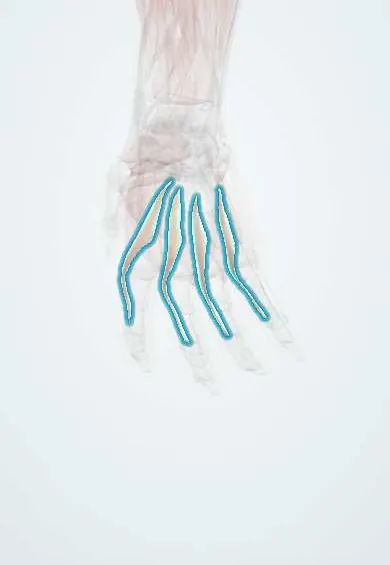
Lumbricals !
The lumbricals are four small palm muscles running from the flexor tendons to the extensor mechanism; they bend the knuckles while straightening the fingers.
Opens in the Durgical app, no sign-up needed.
What it is
Four small worm-shaped muscles in the palm, starting on the FDP tendons.
What it does
Bend the knuckles while straightening the finger joints (the 'tabletop' position).
Key facts
- Origin
- Tendons of FDP
- Insertion
- Lateral sides of the extensor expansions of digits 2–5
- Action
- Flex the MCP joints and extend the IP joints
- Nerve
- 1st–2nd: median nerve; 3rd–4th: deep ulnar nerve (C8–T1)
- Blood supply
- Superficial and deep palmar arches
Relations
- Superficial
- FDS tendons, the superficial palmar arch and the common palmar digital nerves and arteries, under the palmar aponeurosis
- Deep
- The interossei, the adductor pollicis (for the first two), the deep palmar arch and the deep branch of the ulnar nerve
- Course
- From the FDP tendons in the palm each runs along the radial side of an MCP joint, palmar to the deep transverse metacarpal ligament, to the radial lateral band of the extensor mechanism
How to find it
Not palpable. In the palm they are found along the radial side of each FDP tendon beyond the carpal tunnel; their combined action is the tabletop position (knuckles bent, finger joints straight).
Examination
- Tabletop hold: the fingers are held with the knuckles bent and the IP joints straight against pressure; weakness of the ring and little fingers with index and middle preserved fits an ulnar nerve lesion
On imaging
Too small to assess routinely. On high-resolution ultrasound and MRI they are thin bellies on the radial side of the FDP tendons in the palm, beside the common digital nerves. They are rarely imaged for their own sake: imaging assesses the FDP tendons and the median and ulnar nerves instead, and MRI shows denervation of the hand muscles as a group.
Clinical notes
- Ulnar paradox: clawing is worse after an ulnar lesion at the wrist than at the elbow, because the ring and little FDP still pull the fingertips into flexion
- Lumbrical-plus finger: after an FDP cut beyond the lumbrical origin, trying to bend the finger pulls through the lumbrical and paradoxically straightens the IP joints
- The hand is splinted in the intrinsic-plus (safe) position, knuckles bent and finger joints straight, to keep the collateral ligaments and volar plates from shortening
Anatomical variants
- The textbook split (median for the first two, ulnar for the last two) is found in only about half to three-fifths of hands; other patterns, often involving the 3rd lumbrical, occur in a sizeable minority
Memory aid
Median nerve muscles of the hand: LOAF (Lateral two Lumbricals, Opponens pollicis, Abductor pollicis brevis, Flexor pollicis brevis).
Related conditions
Test yourself
Think of the answer, then tap the question to see it.
What is unique about the attachments of the lumbricals?
They are the only muscles that arise from a tendon (FDP) and insert into a tendon (the extensor mechanism).
What is the nerve supply of the lumbricals?
1st and 2nd: median nerve; 3rd and 4th: deep branch of the ulnar nerve.
Why does an ulnar claw affect the ring and little fingers but spare the index and middle?
The 3rd and 4th lumbricals (ulnar) are lost with the interossei, while the median-supplied 1st and 2nd still work.
What is a lumbrical-plus finger?
After an FDP cut beyond the lumbrical origin, trying to bend the finger pulls through the lumbrical and paradoxically straightens the IP joints.
Educational information, not medical advice. For any symptom, see a health professional.
Its names
- Official Latin (TA2)
- musculi lumbricales manus TA2 2532
- Official Spanish (DeCS)
- Músculos lumbricales de la mano
- English (TA2)
- Lumbricals
Related
- Median nerve
- Ulnar nerve in the hand
- Superficial palmar arch
- 2nd metacarpal
- 3rd metacarpal
- 4th metacarpal
- Proximal phalanx, index finger
- Proximal phalanx, middle finger
Sources: Terminologia Anatomica (TA2); Anatomy, Shoulder and Upper Limb, Hand Lumbrical Muscles.
Spotted a mistake?We are always improving for the community. Tell us what is wrong and our team will review it.Report a mistake
Representative model, for educational purposes only.