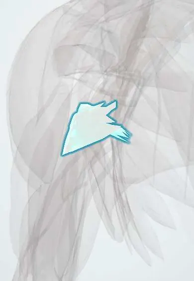
Pec major tendon !
The pectoralis major tendon attaches the large chest muscle to the upper humerus; it can tear during heavy lifts such as the bench press.
Official name: Pectoralis major tendon
Opens in the Durgical app, no sign-up needed.
What it is
The twisted flat tendon where the chest muscle attaches to the front of the humerus.
What it does
Passes the pull of the chest muscle to the arm.
Key facts
- Attaches
- Two-layered, twisted tendon to the lateral lip of the intertubercular groove: the clavicular layer in front and lower, the sternocostal layer behind and higher
- Function
- Adduction and medial rotation of the arm; forms the anterior axillary fold; tears at or near the insertion in heavy pressing
- Tight in
- Abduction with lateral rotation and extension
Relations
- Course
- Fibres from the clavicle, sternum and costal cartilages converge across the front of the axilla into a tendon that twists, so the lowest fibres insert highest on the humerus; this forms the anterior axillary fold
- Deep
- Long biceps tendon in the intertubercular groove, the short head of the biceps and the coracobrachialis; above, the pectoralis minor and clavipectoral fascia
- Lateral
- Anterior deltoid, separated by the deltopectoral groove with the cephalic vein
- Below
- Latissimus dorsi and teres major tendons, inserting on the floor and medial lip of the same groove
- Superficial
- Skin and fat of the anterior axillary fold
How to find it
With the hands pressed together in front of the chest or pushed on the hips, the anterior axillary fold tightens; the tendon is felt as a firm edge at the front of the armpit, running to the upper humerus under the anterior deltoid.
Examination
- Resisted adduction and medial rotation (hands pressed together or on the hips): a gap in the axillary fold, asymmetry and a medial bulge of muscle suggest a tear
- Comparing both anterior axillary folds with the arms abducted shows thinning or loss of the fold on the torn side
On imaging
MRI with axial and oblique coronal images of the chest wall and upper arm shows which part is torn, where (insertion, junction or muscle) and how far it has retracted; ultrasound is useful early. X-rays are usually normal.
Clinical notes
- Complete insertion tears in active people are usually repaired early; chronic tears may need a tendon graft
- Subpectoral biceps tenodesis is done at the lower border of its tendon; the musculocutaneous nerve lies medially and is protected
- A pectoralis major transfer can replace an irreparable subscapularis tendon
- The deltopectoral interval between it and the deltoid, with the cephalic vein, is the main anterior approach to the shoulder
Anatomical variants
- The attachments and the separation between its parts vary
- The sternocostal part may be absent, typically on one side, in Poland syndrome
Memory aid
'A lady between two majors': latissimus dorsi in the floor of the groove, between pectoralis major on the lateral lip and teres major on the medial lip.
Test yourself
Think of the answer, then tap the question to see it.
Where does the pectoralis major tendon insert, and how are its layers arranged?
On the lateral lip of the intertubercular groove; twisted, with the clavicular layer in front and lower, the sternocostal behind and higher.
When does the pectoralis major tendon typically tear?
During the bench press, as the bar is lowered with the arm extended and abducted, at the insertion or musculotendinous junction.
Which part of the pectoralis major tendon tears most, and why can it be missed?
The sternocostal part; an intact clavicular part can keep a near-normal axillary fold.
What signs indicate a pectoralis major tendon tear?
Loss of the anterior axillary fold and a bulge of retracted muscle on the chest during resisted adduction.
Educational information, not medical advice. For any symptom, see a health professional.
Its names
- Official Latin (TA2)
- tendo musculi pectoralis majoris TA2 2301
- Official Spanish (DeCS)
- Tendón del músculo pectoral mayor
- English (TA2)
- Pectoralis major tendon
Related
Sources: Terminologia Anatomica (TA2); Anatomy, Shoulder and Upper Limb, Pectoral Muscles.
Spotted a mistake?We are always improving for the community. Tell us what is wrong and our team will review it.Report a mistake
Representative model, for educational purposes only.