Conditions · Pediatrics and neonatology
Cerebral palsy !
Cerebral palsy is a group of lifelong movement and posture disorders caused by damage to the developing brain. It does not get worse, but needs ongoing therapy.
Updated 2026-11-01
Structures involved
Tap one to see what it is and open it in 3D.
In embryology
In the clinical exam
Why it happens
- Watershed areas and the organs most sensitive to ischemia
- Upper versus lower motor neuron lesions
- Motor cortex and the corticospinal tract
- Basal ganglia and movement control
What it is
A group of permanent disorders of movement and posture caused by a non-progressive injury or malformation of the developing fetal or infant brain. It is the most common cause of physical disability in childhood.
Who gets it
Occurs in about 2 per 1,000 live births worldwide. Prematurity and low birth weight are the strongest associations; in Colombia and Latin America, neonatal infections, perinatal asphyxia, kernicterus and limited access to early intervention also contribute.
Causes
- Most cases (about 85–90%) are congenital: the injury happens before or during birth, often without a single identifiable cause
- Periventricular leukomalacia and intraventricular haemorrhage in preterm infants
- Perinatal hypoxic-ischaemic encephalopathy (a minority of cases)
- Congenital infections (cytomegalovirus, toxoplasmosis, rubella, Zika) and brain malformations
- Kernicterus from severe neonatal hyperbilirubinaemia
- Acquired after the neonatal period: meningitis, head injury (including abusive head trauma), near-drowning
Risk factors
- Preterm birth and low birth weight
- Multiple pregnancy
- Maternal infection, chorioamnionitis or thyroid disease
- Neonatal stroke, seizures or severe jaundice
Pathophysiology
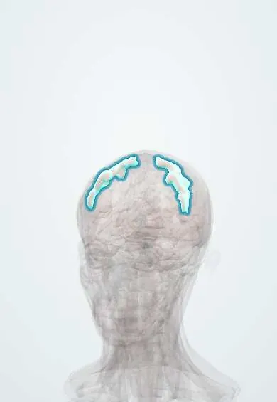
Damage to motor pathways in the immature brain disrupts control of tone and movement. Injury to the corticospinal tracts (motor cortex and periventricular white matter) produces spasticity; injury to the basal ganglia gives dyskinetic forms; injury to the cerebellum gives ataxia. The lesion itself does not progress, but muscle contractures, hip dislocation and scoliosis develop with growth.
Classification
- Spastic (about 80%): hemiplegia, diplegia (legs more than arms, typical of prematurity) or quadriplegia
- Dyskinetic: dystonia or choreoathetosis (basal ganglia, classically kernicterus or severe asphyxia)
- Ataxic: poor balance and coordination (cerebellum)
- Mixed forms
- Severity by the Gross Motor Function Classification System (GMFCS levels I–V)
Symptoms and signs
- Delayed motor milestones (not sitting, crawling or walking at expected ages)
- Increased tone with brisk reflexes, clonus and persistent primitive reflexes; or low tone in early infancy
- Early hand preference before 12 months (suggests hemiplegia)
- Toe walking or scissoring gait
- Feeding and swallowing problems, drooling
- Associated conditions: epilepsy, intellectual disability, visual and hearing impairment, speech disorders
Diagnosis
- Clinical
- A clinical diagnosis based on history (pregnancy, birth, neonatal course) and a neurological examination of tone, reflexes, posture and milestones. Standardised tools such as the Hammersmith Infant Neurological Examination and General Movements Assessment allow diagnosis or high-risk designation in early infancy.
- Tests
- Metabolic and genetic testing when there is no clear perinatal cause, a family history or regression. EEG if seizures are suspected; hearing and vision assessment.
- Imaging
- Brain MRI is the imaging of choice and shows the lesion in most children (periventricular white matter injury, basal ganglia or cortical lesions, malformations); cranial ultrasound in preterm newborns. Hip radiographs for surveillance.
Differential diagnosis
- Neurodegenerative and metabolic disorders (leukodystrophies, mitochondrial disease)
- Spinal muscular atrophy and muscular dystrophies
- Hereditary spastic paraplegia
- Spinal cord lesions or tumours
Goal of treatment
Maximise function, independence, communication and participation, prevent contractures and hip dislocation, and treat associated problems, through a multidisciplinary team that supports the family.
Treatment
- Rehabilitation
- Early intervention with physiotherapy, occupational therapy and speech therapy; orthoses, mobility aids and communication devices.
- Medical
- Spasticity treatment with oral muscle relaxants (e.g. baclofen), botulinum toxin injections to selected muscles or intrathecal baclofen; antiseizure drugs for epilepsy.
- Surgical
- Orthopaedic surgery (tendon lengthening, hip and spine procedures) and, in selected children, selective dorsal rhizotomy.
- Follow-up
- Nursing priorities: nutrition and safe swallowing, aspiration prevention, skin care and repositioning, hip and spine surveillance, and support for caregivers.
Complications
- Muscle contractures and joint deformities
- Hip subluxation and dislocation; scoliosis
- Aspiration pneumonia and malnutrition
- Epilepsy
- Osteopenia and fractures
- Pain, pressure injuries and constipation
Prognosis
Lifelong but non-progressive. Most children with mild forms walk and attend regular school; survival is shortest in those with severe motor impairment, feeding problems and epilepsy, mainly from respiratory causes. Function at age 2 helps predict walking ability.
Prevention
- Good prenatal care and prevention of preterm birth
- Antenatal magnesium sulphate for women at risk of very preterm birth (neuroprotection)
- Therapeutic hypothermia for term newborns with moderate to severe hypoxic-ischaemic encephalopathy
- Prevention and treatment of severe neonatal jaundice; rubella vaccination
Test yourself
Think of the answer, then tap the question to see it.
What is the most common type of cerebral palsy?
Spastic cerebral palsy, about 80% of cases.
Which type of cerebral palsy is typical of preterm infants with periventricular leukomalacia?
Spastic diplegia, with the legs more affected than the arms.
Which finding suggests a diagnosis other than cerebral palsy?
Loss of acquired skills (regression), since cerebral palsy is non-progressive.
What is the imaging study of choice in suspected cerebral palsy?
Brain MRI.
Which neonatal condition causes dyskinetic cerebral palsy through basal ganglia injury?
Kernicterus from severe hyperbilirubinaemia (also severe hypoxic-ischaemic injury).
Related conditions
- Epilepsy and seizures
- Hydrocephalus
- Bacterial meningitis
- Scoliosis
- Hip dislocation
- Strabismus and amblyopia
- Neonatal jaundice
Educational information, not medical advice. For any symptom, see a health professional.
Sources: About Cerebral Palsy (Centers for Disease Control and Prevention (CDC), 2026).
Spotted a mistake?We are always improving for the community. Tell us what is wrong and our team will review it.Report a mistake
Representative model, for educational purposes only.