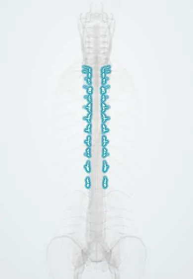
Rib-to-spine ligaments !
The costovertebral ligaments are small ligaments joining each rib to the thoracic vertebrae; they let the ribs lift with each breath.
Official name: Radiate ligament of head of rib
Opens in the Durgical app, no sign-up needed.
What it is
Small ligaments joining the head and neck of each rib to the thoracic vertebrae.
What it does
Hold the ribs to the spine while letting them lift with each breath.
Key facts
- Attaches
- Radiate: rib head to bodies above and below and disc; intra-articular: head crest to disc; costotransverse (superior, lateral, of neck): rib neck and tubercle to transverse processes
- Function
- Bind ribs to thoracic vertebrae while allowing rib elevation in breathing (pump-handle upper ribs, bucket-handle lower ribs)
Relations
- In front
- Sympathetic trunk lying on the rib heads, then the parietal pleura
- Course
- The ventral ramus (intercostal nerve) and the intercostal vessels pass in front of the superior costotransverse ligament; the dorsal ramus passes behind it
- Behind
- Erector spinae and the levatores costarum over the costotransverse joints
- Medial
- The intervertebral disc and foramen: the rib head sits at the disc level, close to the exiting thoracic nerve
How to find it
In dissection, from the front after the pleura is lifted: the radiate ligament fans from the rib head onto the two bodies and the disc. From behind, deep to the erector spinae and levatores costarum: the costotransverse ligaments join the neck and tubercle of the rib to the transverse process.
Examination
- Chest expansion: a tape at about the 4th intercostal space measures the chest from full breath out to full breath in; a small difference fits stiff costovertebral joints, as in ankylosing spondylitis
On imaging
CT shows the joints of the rib heads and the costotransverse joints best: erosions or bony fusion in ankylosing spondylitis, spurs in osteoarthritis. MRI can show early inflammation (bone marrow oedema) at these joints.
Clinical notes
- Costotransversectomy removes the rib head and transverse process to reach thoracic discs and vertebral bodies from behind and to the side
- In thoracic surgery the rib head is a level landmark: it leads to the disc above its own vertebra (the 6th rib head to the T5–T6 disc)
- Pain from these joints can mimic thoracic facet, cardiac or pleural pain
Development
Ribs start as the costal parts of the thoracic vertebral cartilage models and separate from them in the embryo (about week 8), leaving joints at the head and tubercle. Elsewhere the costal part fuses with the vertebra; if it stays free at C7 or L1 it forms a cervical or lumbar rib.
Related conditions
Test yourself
Think of the answer, then tap the question to see it.
Which ribs meet two vertebral bodies and the disc between them through the rib-to-spine ligaments?
Ribs 2–9; ribs 1, 10, 11 and 12 usually meet only one body.
Which joint do ribs 11 and 12 lack, so that they move more freely?
The costotransverse joint: only the rib head joins the vertebra.
What happens to the costovertebral joints in ankylosing spondylitis, and how is it measured?
They fuse, so chest expansion falls, a classic exam measurement.
In thoracic surgery, how is the rib head used as a level landmark?
It leads to the disc above its own vertebra: the 6th rib head points to the T5–T6 disc.
Educational information, not medical advice. For any symptom, see a health professional.
Its names
- Official Latin (TA2)
- ligamentum radiatum capitis costae; ligamentum intraarticulare capitis costae; ligamentum costotransversarium TA2 1722
- Official Spanish (DeCS)
- Ligamentos costovertebrales y costotransversos
- English (TA2)
- Radiate ligament of head of rib
Related
- Mid-back facet joints
- C7 vertebra
- T1 vertebra
- T10 vertebra
- T11 vertebra
- T12 vertebra
- T2 vertebra
- T3 vertebra
Sources: Terminologia Anatomica (TA2); Anatomy, Thorax, Ribs; Anatomy of the Human Body (20th ed.): Costovertebral Articulations (Lea & Febiger (Gray's Anatomy, 1918), 1918) · adapted, Public domain; 7.4 The Thoracic Cage (OpenStax (Rice University), 2022) · adapted, CC BY 4.0; 7.5 Embryonic Development of the Axial Skeleton (OpenStax (Rice University), 2022) · adapted, CC BY 4.0.
Spotted a mistake?We are always improving for the community. Tell us what is wrong and our team will review it.Report a mistake
Representative model, for educational purposes only.