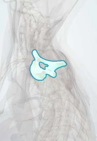
T1 vertebra !
T1 is the first thoracic (upper back) vertebra, just below the neck; it carries the first pair of ribs.
Official name: T1
Opens in the Durgical app, no sign-up needed.
What it is
The first mid-back vertebra, which carries the first ribs.
What it does
Holds up the trunk and, with the ribs and breastbone, forms the rib cage, which protects the heart and lungs. The ribs make this part of the spine stiff.
Key facts
- Joints with
- C7 above and T2 below (discs, facet joints); head of rib 1 (full facet) and upper half of head of rib 2; tubercle of rib 1
- Landmarks
- Full superior costal facet for rib 1 plus inferior demifacet for rib 2; cervical-like body; long, nearly horizontal spinous process, prominent like C7
Relations
- In front
- Longus colli, then the esophagus with the trachea in front of it; the thoracic duct climbs on the left side of the esophagus
- Lateral
- Head of the first rib on its full facet; over the neck of the first rib, from medial to lateral: the sympathetic trunk (stellate ganglion), the superior intercostal artery and the T1 ventral ramus rising to the brachial plexus
- Above
- The dome of the pleura and the lung apex rise above the first rib on each side, in front of the neck of the rib
- Behind
- Long, prominent spine with trapezius, rhomboids, splenius and serratus posterior (superior) attached
How to find it
With the neck flexed, C7 and T1 are the two largest bumps at the base of the neck. When the head is turned side to side, the spine that moves slightly is C7 and the one that stays still is T1. The superior angle of the scapula lies about one level lower.
Examination
- T1 root: weak spreading of the fingers (interossei) and numbness of the inner arm just above the elbow; with the C8 findings this places the lesion at the lower trunk or the C8–T1 roots
- Horner syndrome check: drooping upper lid, a smaller pupil (difference larger in dim light) and less sweating on that side of the face; with arm signs it suggests disease at the lung apex or a root avulsion near T1
On imaging
On an AP film T1 is recognised by its upward-pointing transverse processes meeting the first ribs. Lateral views are blocked by the shoulders, so CT is used after trauma. CT or MRI of the apex shows a Pancoast tumor and its spread to the roots and chest wall.
Common problems
- Compression fractures from osteoporosis, sometimes after a minor fall or even a cough.
- A rounded upper back (kyphosis) from posture, wedge fractures or Scheuermann disease in teens.
- Scoliosis: a sideways curve with rotation, often starting in adolescence.
How it is diagnosed
X-rays for fractures and curves (scoliosis is measured with the Cobb angle). MRI tells new fractures from old ones. A bone density scan (DEXA) checks for osteoporosis.
Treatment
- Osteoporosis fractures: pain control, a short time in a brace, and treatment of the osteoporosis.
- Painful fractures that do not settle: kyphoplasty or vertebroplasty (cement injected into the bone).
- Large or progressive curves: bracing in growing children, sometimes fusion surgery.
Clinical notes
- Klumpke palsy (lower trunk, C8–T1), from a forceful upward pull on the arm, as in a breech delivery or grabbing something while falling: claw hand, sometimes with Horner syndrome
- The stellate ganglion lies in front of the C7 transverse process and the neck of the first rib, the target of blocks for some pain syndromes of the arm
- Fractures this high need great force, because the ribs and sternum brace the segment; associated chest and great-vessel injuries are looked for
- Surgery for thoracic outlet syndrome often removes the first rib, whose head meets T1 alone, together with any cervical rib or fibrous band, taking care of the lower trunk and the subclavian vessels that cross it
Development
Ribs start as the costal parts of the cartilage model of each thoracic vertebra and separate from it by about the eighth week; T1 carries the full facet for the first rib. In the neck the same element stays fused within the transverse process.
Test yourself
Think of the answer, then tap the question to see it.
Which features does a Pancoast tumor next to the T1 vertebra cause, and why?
Shoulder and arm pain, weak hand muscles and Horner syndrome, because it invades the C8 and T1 roots and the sympathetic trunk.
Which ribs articulate with the body of the T1 vertebra?
The first rib on a full costal facet and the upper half of the head of the second rib on an inferior demifacet.
What is Klumpke palsy, which involves the T1 root?
A lower trunk (C8–T1) injury from a forceful upward pull on the arm, as in a breech delivery: claw hand, sometimes with Horner syndrome.
From medial to lateral, which structures cross the neck of the first rib beside the T1 vertebra?
The sympathetic trunk (stellate ganglion), the superior intercostal artery and the T1 ventral ramus rising to the brachial plexus.
Educational information, not medical advice. For any symptom, see a health professional.
Its names
- Official Latin (TA2)
- vertebra thoracica I TA2 1059
- Official Spanish (DeCS)
- Vértebra dorsal D1 DeCS D013904
- English (TA2)
- T1
Related
- C7–T1 disc
- T1–T2 disc
- Posterior intercostal arteries
- Anterior longitudinal ligament
- Rib-to-spine ligaments
- Dura (dural sac)
- Endplates
- Neck facet joints
Sources: Terminologia Anatomica (TA2); DeCS; Anatomy, Back, Thoracic Vertebrae; 7.5 Embryonic Development of the Axial Skeleton (OpenStax (Rice University), 2022) · adapted, CC BY 4.0.
Spotted a mistake?We are always improving for the community. Tell us what is wrong and our team will review it.Report a mistake
Representative model, for educational purposes only.