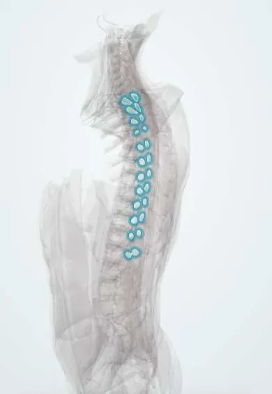
Mid-back facet joints !
The thoracic facet joints are pairs of small, nearly upright sliding joints at the back of the mid-back; they allow turning but little bending.
Official name: Zygapophysial (facet) joints
Opens in the Durgical app, no sign-up needed.
What it is
Pairs of small sliding joints at the back of the mid-back, set almost upright, which allow turning but little bending.
What it does
Guide and limit movement (lumbar facets block twisting), and share the load with the disc, especially when you lean back.
Key facts
- Where
- Between articular processes from T1–T2 to T12–L1 (zygapophysial joints)
- Function
- Synovial plane joints; facets near the coronal plane, allow rotation and side bending, limit flexion-extension (ribs limit further); nerve: medial branches of dorsal rami
Relations
- Lateral
- The costotransverse joints with the rib tubercles (T1–T10), just outside each facet
- In front
- The intervertebral foramen with the thoracic spinal nerve; in front of that, the rib head at the costovertebral joint
- Behind
- Multifidus, rotatores, semispinalis and the erector spinae; the medial branches of the dorsal rami cross near the upper outer corner of the transverse process
- Medial
- Laminae and ligamentum flavum; inside them, the thoracic spinal cord in a relatively narrow canal
How to find it
The thoracic spinous processes slant steeply downward, so in the mid-thoracic region the palpable tip lies near the level of the vertebra below; the facets and transverse processes of a vertebra therefore sit higher than its tip, a few centimetres to each side of the midline.
Examination
- Prone springing (posteroanterior pressure) over the transverse processes: reproduction of the usual local pain suggests a facet or costotransverse source; it does not tell the two apart
On imaging
Overlapping ribs hide them on plain radiographs. CT shows arthrosis and, after trauma, widened or perched facets in flexion-distraction injuries. Fat-suppressed MRI can show joint fluid or bone oedema around an inflamed facet.
Common problems
- Mid-back pain from strain or arthritis.
How it is diagnosed
There is no single sign. CT or MRI show arthritis; a numbing injection of the nerves to the joint (medial branch block) shows whether the joint is the pain source.
Treatment
- Physical therapy and medicine; injections when needed.
Clinical notes
- Flexion-distraction (Chance-type) injuries at the thoracolumbar junction tear the facet capsules and the posterior ligament complex; facet widening on CT signals instability
- Ankylosing spondylitis and DISH can fuse these joints; a fused thoracic spine breaks like a long bone after minor trauma
- Thoracic facet pain is confirmed with medial branch blocks, but the nerve's path over the transverse process varies more than in the lumbar spine
Anatomical variants
- The level where the facets change from coronal to sagittal varies, usually at T11–T12 or T12–L1, and it may differ between left and right
Test yourself
Think of the answer, then tap the question to see it.
Which movements do the thoracic facet joints allow and limit?
Their facets lie near the coronal plane, so they allow rotation and side bending and limit flexion-extension (the ribs limit movement further).
Why do spine fractures concentrate at T11–L2?
At the thoracolumbar junction the facets turn from coronal to sagittal, so stress concentrates there; it is the most common site of spine fractures.
Why does a fused thoracic spine in ankylosing spondylitis or DISH need special care after minor trauma?
These diseases can fuse the thoracic facet joints, and a fused thoracic spine breaks like a long bone even after minor trauma.
Educational information, not medical advice. For any symptom, see a health professional.
Its names
- Official Latin (TA2)
- articulationes zygapophysiales TA2 1707
- Official Spanish (DeCS)
- Articulación cigapofisaria DeCS D021801
- English (TA2)
- Zygapophysial (facet) joints
- Also called
- facet joints · juncturae zygapophyseales · zygapophysial joints
Related
- Rib-to-spine ligaments
- L1 vertebra
- T1 vertebra
- T10 vertebra
- T11 vertebra
- T12 vertebra
- T2 vertebra
- T3 vertebra
Sources: Terminologia Anatomica (TA2); DeCS; Anatomy, Back, Thoracic Vertebrae; Anatomy of the Human Body (20th ed.): Surface Anatomy of the Back (Lea & Febiger (Gray's Anatomy, 1918), 1918) · adapted, Public domain.
Spotted a mistake?We are always improving for the community. Tell us what is wrong and our team will review it.Report a mistake
Representative model, for educational purposes only.