Brugada syndrome !
An inherited electrical heart disorder with a typical ECG pattern that can cause fainting or sudden death, often at rest or during sleep.
Updated 2026-11-01
Structures involved
Tap one to see what it is and open it in 3D.
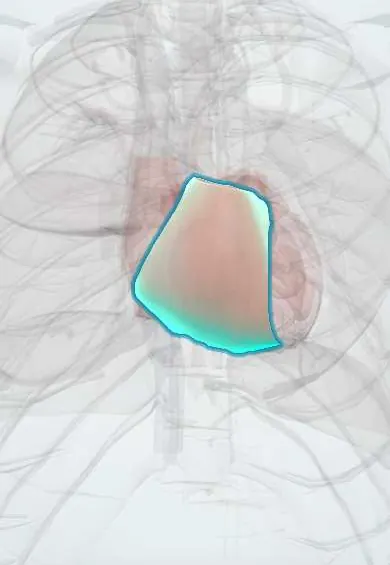
- Right ventricle
- Bundle branches
- Pulmonary trunk
- Bundle of His
- Sinoatrial (SA) node
- Atrioventricular (AV) node
Why it happens
- Mendelian inheritance: autosomal dominant, recessive and X-linked
- The ventricular action potential
- Ion channels and gating
- How arrhythmias arise
What it is
Brugada syndrome is an inherited channelopathy, a disease of cardiac ion channels in a structurally normal heart. It produces a characteristic coved ST-segment elevation in the right precordial leads (V1 and V2) and predisposes to polymorphic ventricular tachycardia and ventricular fibrillation, causing syncope or sudden death, typically at rest, during sleep or with fever.
Who gets it
Estimated at about 5 in 10,000 people worldwide, much more common in Southeast Asia, where it is linked to sudden unexplained nocturnal death. Symptoms appear most often in men in their thirties and forties; men are affected about 8 to 10 times more often. No specific Colombian data exist.
Causes
- Loss-of-function variants in SCN5A, the cardiac sodium channel gene, in about a fifth to a third of cases
- Variants in other channel genes (calcium channel and others) in a small minority; in many no gene is found
- Autosomal dominant inheritance with incomplete penetrance; some cases are new mutations
- Unmasking factors: fever, sodium channel blockers (class I antiarrhythmics), tricyclic antidepressants, lithium, cocaine, alcohol, large meals, hypokalaemia
Risk factors
- Family history of Brugada syndrome or of sudden death in a young relative
- Male sex (testosterone influences the outward potassium current)
- Southeast Asian ancestry
- Fever, especially in children
- Use of drugs that block sodium channels
Pathophysiology
Reduced inward sodium current, mostly in the right ventricular outflow tract epicardium, leaves the transient outward potassium current unopposed early in the action potential. This creates a voltage gradient between epicardium and endocardium, seen as ST elevation in V1 and V2. Heterogeneous loss of the action potential dome allows phase 2 re-entry, which triggers polymorphic ventricular tachycardia and fibrillation. Conduction slowing in the outflow tract contributes. Fever and vagal tone at night accentuate the defect.
Classification
- Type 1 (diagnostic) pattern: coved ST elevation of at least 2 mm followed by a negative T wave in V1 or V2
- Type 2 pattern: saddleback ST elevation, not diagnostic on its own
- Spontaneous versus drug- or fever-induced type 1 pattern
- Symptomatic (syncope, arrest) versus asymptomatic
Symptoms and signs
- Many patients have no symptoms and are found by ECG or family screening
- Syncope, often at rest, at night or with fever
- Agonal breathing or nocturnal gasping during sleep
- Palpitations; atrial fibrillation is more frequent
- Sudden cardiac arrest as the first event
Diagnosis
- Clinical
- Diagnosis rests on a type 1 ECG pattern, spontaneous or after a provocation test, especially with syncope, arrhythmia or family history. Ask about unmasking drugs, fever and family history of sudden death.
- Tests
- 12-lead ECG, including leads placed one or two intercostal spaces higher (second and third spaces) to increase sensitivity. Sodium channel blocker challenge (for example ajmaline or flecainide) in a monitored setting when the pattern is suspected but not spontaneous. Electrolytes, genetic testing (SCN5A) and family screening. Electrophysiological study in selected cases for risk stratification.
- Imaging
- Echocardiography and, when needed, cardiac magnetic resonance to exclude structural disease such as ARVC.
Differential diagnosis
- Arrhythmogenic right ventricular cardiomyopathy
- Right bundle branch block and athlete's heart
- Acute myocardial infarction or pericarditis
- Brugada phenocopies (hyperkalaemia, hypercalcaemia, drug toxicity such as tricyclic antidepressants)
- Long QT syndrome
- Epileptic seizure (nocturnal events)
Goal of treatment
Prevent sudden death, avoid triggers and identify affected relatives.
Treatment
- Procedure
- Implantable cardioverter-defibrillator for survivors of cardiac arrest, documented sustained ventricular arrhythmia, or arrhythmic syncope with a spontaneous type 1 pattern.
- Lifestyle
- Avoid drugs that unmask the pattern (sodium channel blockers, some psychotropics and anaesthetics), cocaine, excess alcohol and large meals; patients consult an updated list of drugs to avoid.
- Acute
- Treat fever promptly with antipyretics and obtain an ECG during febrile illness; electrical storm is treated with isoproterenol infusion in hospital.
- Medical
- Quinidine, which blocks the transient outward current, for recurrent arrhythmias or shocks, or when a defibrillator is not possible.
- Procedure
- Epicardial catheter ablation of the right ventricular outflow tract substrate in patients with recurrent ventricular fibrillation.
- Follow-up
- Nursing: continuous ECG monitoring in hospital, fever control, review of every medication against the list of drugs to avoid, defibrillator education, and family screening.
Complications
- Sudden cardiac death
- Recurrent ventricular fibrillation and electrical storm
- Atrial fibrillation
- Inappropriate defibrillator shocks and device complications
Prognosis
Risk depends on presentation: highest after a cardiac arrest, intermediate with arrhythmic syncope and a spontaneous type 1 pattern, and low in asymptomatic people with only a drug-induced pattern. With a defibrillator and trigger avoidance, high-risk patients can live long lives.
Prevention
- ECG screening of first-degree relatives and genetic testing when a variant is known
- Avoidance of unmasking drugs and prompt treatment of fever
- Anaesthetic and medication planning in known patients
Test yourself
Think of the answer, then tap the question to see it.
Which gene is most often involved in Brugada syndrome?
SCN5A, which encodes the cardiac sodium channel.
What is the diagnostic (type 1) ECG pattern?
Coved ST elevation of at least 2 mm with a negative T wave in V1 or V2.
Which common situation can unmask the pattern and trigger arrhythmia?
Fever (also sodium channel blockers, tricyclics, cocaine and alcohol).
What is the only proven therapy to prevent sudden death in high-risk patients?
An implantable cardioverter-defibrillator.
When do arrhythmic events typically occur?
At rest or during sleep, when vagal tone is high.
Related conditions
- Syncope (vasovagal, orthostatic hypotension, cardiac)
- Cardiac arrest (VF, pulseless VT, asystole, PEA)
- Ventricular tachycardia
- Arrhythmogenic right ventricular cardiomyopathy
- Long QT syndrome and torsades de pointes
- Atrial fibrillation
- Sudden infant death syndrome (SIDS)
- Epilepsy and seizures
- Cocaine and sympathomimetic toxicity
- Anticholinergic toxicity (incl. scopolamine «burundanga»)
- Lithium toxicity
- Myocardial Infarction (Heart Attack)
- Acute pericarditis
Educational information, not medical advice. For any symptom, see a health professional.
Sources: Brugada syndrome: MedlinePlus Genetics (MedlinePlus Genetics, US National Library of Medicine, 2026).
Spotted a mistake?We are always improving for the community. Tell us what is wrong and our team will review it.Report a mistake
Representative model, for educational purposes only.