Splenic injury !
Splenic injury is damage to the spleen, usually from a blow to the belly or a fall; it can cause life-threatening bleeding inside the abdomen.
Updated 2026-10-29
Structures involved
Tap one to see what it is and open it in 3D.
In immunology
In histology
In embryology
In physiology
Why it happens
- Hemorrhage: petechiae, purpura, ecchymosis, hematoma, and what blood loss does
- Shock: types, stages and the organs it damages
What it is
Damage to the spleen, from a small capsular tear to complete shattering or loss of its blood supply, usually from blunt abdominal trauma. It can cause life-threatening bleeding into the abdomen.
Who gets it
The spleen is one of the most frequently injured abdominal organs in blunt trauma, mostly from road traffic crashes, falls, assaults and sports. Spontaneous rupture can occur in an enlarged spleen, for example from malaria or infectious mononucleosis.
Causes
- Blunt trauma: road traffic crashes, falls, assaults, contact sports
- Penetrating trauma (stab or gunshot wounds) to the left upper quadrant or lower left chest
- Iatrogenic injury during abdominal surgery or colonoscopy
- Minor trauma or spontaneous rupture of a diseased, enlarged spleen
Risk factors
- Splenomegaly (malaria, infectious mononucleosis, haematological disease, portal hypertension)
- Fractures of the lower left ribs
- Motorcycle use and high-energy trauma
- Anticoagulation
Pathophysiology
The spleen is soft, highly vascular and fixed under the lower left ribs, so it tears easily with blunt force. Bleeding may be contained under the capsule (subcapsular haematoma) or flow freely into the peritoneum. Blood under the left diaphragm irritates the phrenic nerve and refers pain to the left shoulder (Kehr sign). A contained haematoma can rupture days later.
Classification
- AAST (American Association for the Surgery of Trauma) organ injury scale, grades I to V, based on haematoma size, laceration depth and vascular injury
- Grade V: shattered spleen or hilar vascular injury that devascularises it
- Haemodynamically stable vs unstable (guides management more than grade)
Symptoms and signs
- Left upper quadrant pain and tenderness
- Left shoulder tip pain (Kehr sign)
- Abdominal distension or peritonism
- Signs of blood loss: tachycardia, pallor, hypotension
- Bruising over the left flank or lower left ribs
Diagnosis
- Clinical
- ATLS primary survey; mechanism of injury, left upper quadrant tenderness, Kehr sign, lower left rib fractures and haemodynamic status.
- Tests
- Serial haemoglobin, blood group and crossmatch, coagulation, lactate and base deficit.
- Imaging
- eFAST detects free fluid at the bedside. In stable patients, contrast CT grades the injury and shows active extravasation (contrast blush) or pseudoaneurysm. Unstable patients with free fluid go to surgery without CT.
Differential diagnosis
- Liver or left kidney injury
- Left rib fractures and haemothorax
- Diaphragmatic injury
- Pancreatic tail injury
- Hollow viscus injury
Goal of treatment
Stop the bleeding and keep the patient alive, preserving the spleen whenever it is safe, and protect asplenic patients against severe infection.
Treatment
- Acute
- ATLS resuscitation: airway, breathing, haemorrhage control, blood products and a massive transfusion protocol when needed.
- Follow-up
- Non-operative management for haemodynamically stable patients, whatever the grade: close observation in a unit able to operate, serial examinations and haemoglobin, and activity restriction afterwards.
- Procedure
- Angioembolisation of the splenic artery or its branches for active contrast extravasation or pseudoaneurysm in stable patients.
- Surgical
- Emergency laparotomy and splenectomy for unstable patients or failed non-operative management; splenorrhaphy or partial splenectomy in selected cases.
- Medical
- After splenectomy or extensive embolisation: vaccines against pneumococcus, meningococcus and Haemophilus influenzae type b, yearly influenza vaccine, and education about early antibiotics for fever.
Complications
- Haemorrhagic shock and death
- Delayed rupture of a subcapsular haematoma
- Failure of non-operative management
- Splenic abscess, infarction or pseudocyst after embolisation
- Overwhelming post-splenectomy infection from encapsulated bacteria
- Pancreatic tail injury or fistula after splenectomy
Prognosis
Most stable patients, including children, are treated successfully without surgery. Outcome depends on associated injuries and the speed of haemorrhage control; the risk of severe infection after splenectomy is lifelong.
Prevention
- Road safety, helmets and seat belts
- Avoidance of contact sports when the spleen is enlarged (e.g., during infectious mononucleosis)
- Vaccination and fever education after splenectomy
Test yourself
Think of the answer, then tap the question to see it.
What is Kehr sign in splenic injury?
Left shoulder tip pain from blood irritating the left hemidiaphragm, referred through the phrenic nerve (C3 to C5).
What decides between operative and non-operative management of splenic injury?
Mainly haemodynamic status: stable patients get non-operative management whatever the grade; unstable ones need surgery.
Which CT finding in a stable patient often leads to splenic angioembolisation?
Active contrast extravasation (blush) or a pseudoaneurysm.
Against which bacteria is a patient vaccinated after splenectomy?
Encapsulated bacteria: Streptococcus pneumoniae, Neisseria meningitidis and Haemophilus influenzae type b.
Which scale grades splenic injuries?
The AAST organ injury scale, grades I to V, from small subcapsular haematoma to shattered or devascularised spleen.
Related conditions
Educational information, not medical advice. For any symptom, see a health professional.
Sources: Splenic Trauma (StatPearls (NCBI Bookshelf), 2025).
Spotted a mistake?We are always improving for the community. Tell us what is wrong and our team will review it.Report a mistake
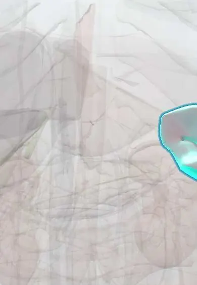
Representative model, for educational purposes only.