Cardiovascular system · Abdomen
Mesenteric veins !
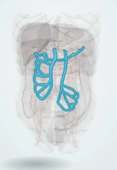
The mesenteric veins collect nutrient-rich blood from the intestines and carry it toward the portal vein and the liver.
Official name: Superior mesenteric vein
Opens in the Durgical app, no sign-up needed.
What it is
The superior and inferior mesenteric veins and their colic and gastro-omental branches.
What it does
Drain the intestines into the portal vein.
Key facts
- Drains
- SMV: small intestine, cecum, ascending and transverse colon, pancreatic head; IMV: descending and sigmoid colon, upper rectum
- Into
- SMV joins splenic vein to form portal vein; IMV into splenic vein (varies: SMV or the confluence)
Relations
- Course
- Superior mesenteric vein: up in the root of the mesentery, across the front of the third part of the duodenum and the uncinate process, to the neck of the pancreas. Inferior mesenteric vein: up from the pelvis on the left psoas major, then behind the body of the pancreas
- Medial
- Superior mesenteric vein: the superior mesenteric artery on its left side
- Behind
- Superior mesenteric vein: third part of the duodenum, uncinate process and inferior vena cava; inferior mesenteric vein: left psoas major, with the left gonadal vessels and the left ureter close by
How to find it
At surgery the superior mesenteric vein is found in the root of the mesentery, to the right of its artery, and followed up under the neck of the pancreas to the portal confluence. The inferior mesenteric vein is found on its own just left of the duodenojejunal flexure, under a peritoneal fold, and is tied there or at the lower border of the pancreas.
On imaging
Contrast CT in the portal venous phase shows both veins; a filling defect means thrombosis. In midgut volvulus the vein and mesentery wrap around the artery (whirlpool sign) on CT or Doppler ultrasound.
Common problems
- Portal hypertension in cirrhosis: blood backs up, causing swollen veins in the esophagus (varices) that can bleed, an enlarged spleen and fluid in the abdomen (ascites).
How it is diagnosed
Ultrasound with Doppler, CT, endoscopy for varices.
Treatment
- Medicines that lower the pressure, banding of varices, a shunt (TIPS), liver transplant.
Clinical notes
- Mesenteric venous thrombosis, usually with a clotting disorder, causes a slower, less obvious bowel ischemia than an arterial blockage
- The superior mesenteric–portal confluence behind the neck of the pancreas is the critical point in pancreatoduodenectomy: tumour contact with it decides resectability
- In left colon and rectal surgery the inferior mesenteric vein is divided near the lower border of the pancreas to free the colon for a tension-free join
Anatomical variants
- The inferior mesenteric vein may end in the splenic vein, the superior mesenteric vein or the confluence of the two (texts differ on which is commonest)
- Tributaries of the superior mesenteric vein, such as the gastrocolic trunk and the jejunal trunks, vary widely and are mapped on CT before pancreatic or colon surgery
Development
They arise from the vitelline veins; the superior mesenteric vein opens into a venous ring around the duodenum that is remodelled into the portal vein. Rotation of the midgut leaves it to the right of the artery, so a reversed position on imaging points to malrotation.
Related conditions
Test yourself
Think of the answer, then tap the question to see it.
On which side of the superior mesenteric artery does the superior mesenteric vein lie?
On the right; a vein to the left of the artery on imaging suggests intestinal malrotation.
Where does the inferior mesenteric vein usually end?
In the splenic vein behind the pancreas (it may end in the superior mesenteric vein or the confluence); it climbs alone, just left of the duodenojejunal flexure.
Which parts of the gut do the superior and inferior mesenteric veins drain?
Superior: small intestine, cecum, ascending and transverse colon, pancreatic head. Inferior: descending and sigmoid colon and upper rectum.
How does mesenteric venous thrombosis differ from arterial blockage?
It causes a slower, less obvious bowel ischemia, usually in a person with a clotting disorder.
Why is the superior mesenteric–portal confluence critical in pancreatoduodenectomy?
Tumour contact with the confluence, behind the neck of the pancreas, decides whether the tumour can be removed.
Educational information, not medical advice. For any symptom, see a health professional.
Its names
- Official Latin (TA2)
- vena mesenterica superior; vena mesenterica inferior TA2 5113
- Official Spanish (DeCS)
- Venas mesentéricas DeCS D008642
- English (TA2)
- Superior mesenteric vein
- Also called
- vena mesenterica cranialis
Related
Sources: Terminologia Anatomica (TA2); DeCS; Anatomy, Abdomen and Pelvis: Superior Mesenteric Vein; Anatomy, Abdomen and Pelvis, Portal Venous System (Hepatic Portal System); Anatomy of the Human Body (20th ed.): Development of the Vascular System (Lea & Febiger (Gray's Anatomy, 1918), 1918) · adapted, Public domain.
Spotted a mistake?We are always improving for the community. Tell us what is wrong and our team will review it.Report a mistake
Representative model, for educational purposes only.