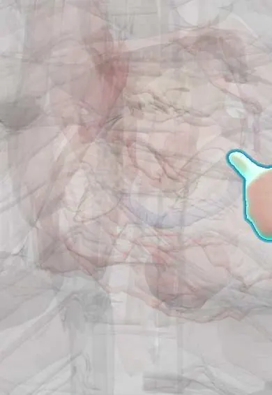
Mechanical complications of myocardial infarction !
Days to weeks after a heart attack, the dead muscle can tear, bulge or inflame the sac around the heart. These are rare but often deadly emergencies.
Updated 2026-11-01
Structures involved
Tap one to see what it is and open it in 3D.
- Left ventricle papillary muscles
- Interventricular septum
- Left ventricle
- Pericardium
- Mitral anterior leaflet
- Mitral posterior leaflet
- Chordae tendineae
- LAD (anterior descending artery)
- Right coronary artery
- Circumflex artery
- Right ventricle
In the clinical exam
Why it happens
- Granulation tissue and scar formation
- The myocardial infarct over time: what the pathologist sees from hours to months
- Reversible vs irreversible cell injury: the point of no return
- Coronary circulation and myocardial ischemia
- Shock physiology
What it is
Structural problems caused by necrosis of the infarcted myocardium: rupture of a papillary muscle (acute mitral regurgitation), rupture of the interventricular septum (ventricular septal defect), rupture of the left ventricular free wall (haemopericardium and tamponade), true or false left ventricular aneurysm, and pericarditis, either early or late (Dressler syndrome).
Who gets it
Uncommon in the era of rapid reperfusion, but still frequent in exam questions and in settings where patients arrive late. In Colombia and Latin America, delayed consultation and limited access to primary angioplasty outside large cities keep late-presenting infarctions, and therefore these complications, relevant. Mortality remains very high when rupture occurs.
Causes
- Transmural infarction with softening of necrotic muscle, typically 3 to 7 days after onset (peak of macrophage activity and lowest tensile strength)
- Papillary muscle rupture: most often the posteromedial papillary muscle, which has a single blood supply (usually from the right coronary or circumflex artery)
- Septal rupture: anterior infarction (left anterior descending artery) or inferior infarction
- Ventricular aneurysm: thin, scarred, dyskinetic wall weeks to months after a large anterior infarction
- Dressler syndrome: immune reaction to cardiac antigens released by the necrotic muscle, weeks after the infarction
Risk factors
- Late presentation or no reperfusion therapy
- First, large, transmural infarction (no previous collaterals)
- Older age and female sex
- Hypertension during the acute phase
- Single-vessel occlusion with poor collateral flow
Pathophysiology
After an infarction, neutrophils and then macrophages clear the dead myocytes; between about day 3 and day 14 the wall is soft before collagen scar forms. Under ventricular pressure the weakened tissue can tear. A torn papillary muscle makes the mitral leaflet flail, producing sudden severe regurgitation and pulmonary oedema. A septal tear creates a left-to-right shunt that overloads the right heart and the lungs. A free-wall tear fills the pericardium with blood and causes tamponade. Later, the mature scar can bulge outward as an aneurysm that pumps poorly, holds thrombus and generates ventricular arrhythmias.
Classification
- Early (hours to about 2 weeks): free-wall rupture, septal rupture, papillary muscle rupture, early (peri-infarction) pericarditis
- Late (weeks to months): true left ventricular aneurysm, pseudoaneurysm (contained rupture), Dressler syndrome
- Non-mechanical complications to keep in mind: arrhythmias, cardiogenic shock, mural thrombus with embolism
Symptoms and signs
- Sudden dyspnoea, pulmonary oedema and hypotension a few days after an infarction
- New holosystolic murmur (apical radiating to the axilla in papillary rupture; left sternal border with a thrill in septal rupture)
- Sudden collapse with distended neck veins and pulseless electrical activity in free-wall rupture
- Pleuritic chest pain relieved by leaning forward, fever and a pericardial friction rub in pericarditis
- Persistent ST elevation weeks after the infarction, heart failure or arrhythmias in a ventricular aneurysm
Diagnosis
- Clinical
- Suspect a mechanical complication in any patient who deteriorates suddenly between about day 3 and day 7 after an infarction, especially with a new murmur, signs of tamponade or shock. Dressler syndrome is suspected with fever, pleuritic pain and a rub weeks after the event.
- Tests
- 12-lead ECG (persistent ST elevation in an aneurysm; diffuse ST elevation and PR depression in pericarditis); troponin and inflammatory markers; right heart catheterisation shows a step-up in oxygen saturation from the right atrium to the right ventricle in septal rupture and large v waves in the wedge pressure in acute mitral regurgitation.
- Imaging
- Bedside echocardiography is the key test: it shows a flail mitral leaflet, a septal defect with left-to-right flow, pericardial effusion with tamponade, or an aneurysm with or without thrombus. Cardiac MRI or CT helps to separate a true aneurysm from a pseudoaneurysm.
Differential diagnosis
- Reinfarction or extension of the infarction
- Cardiogenic shock from pump failure without rupture
- Pulmonary embolism
- Aortic dissection
- Acute pericarditis of other causes
Goal of treatment
Stabilise circulation, reduce afterload and correct the mechanical defect early with surgery or a catheter procedure, while treating the underlying coronary disease; treat pericarditis to relieve pain and prevent recurrence.
Treatment
- Acute
- Haemodynamic support: vasodilators to reduce afterload when blood pressure allows, inotropes, and mechanical circulatory support as a bridge to repair.
- Acute
- Free-wall rupture with tamponade: urgent pericardiocentesis as a temporary measure and emergency surgery.
- Surgical
- Papillary muscle rupture: urgent mitral valve repair or replacement, often with coronary bypass grafting.
- Surgical
- Septal rupture: surgical or transcatheter closure of the defect.
- Medical
- Left ventricular aneurysm: heart failure therapy, anticoagulation when a mural thrombus is present, and an implantable defibrillator or surgery for refractory arrhythmias or heart failure.
- Medical
- Post-infarction and Dressler pericarditis: aspirin and colchicine; non-steroidal anti-inflammatory drugs other than aspirin and corticosteroids are avoided early after infarction because they may impair healing of the scar.
- Follow-up
- Nursing priorities: continuous ECG and blood pressure monitoring, daily auscultation for new murmurs or rubs in the first week, urine output, oxygen saturation and early report of sudden deterioration.
Complications
- Cardiogenic shock and death
- Cardiac tamponade
- Acute pulmonary oedema
- Mural thrombus with stroke or systemic embolism
- Ventricular tachycardia and sudden death
- Recurrent pericarditis
Prognosis
Rupture of the free wall, septum or papillary muscle carries very high mortality without urgent repair, and remains high even with surgery. Ventricular aneurysm worsens long-term survival through heart failure and arrhythmias. Dressler syndrome usually responds to anti-inflammatory treatment but can recur.
Prevention
- Early reperfusion of acute myocardial infarction (primary angioplasty or fibrinolysis)
- Recognising symptoms of a heart attack and calling for help without delay
- Blood pressure control during the acute phase
- Neurohormonal therapy after a large infarction to limit ventricular remodelling
Test yourself
Think of the answer, then tap the question to see it.
A patient develops sudden pulmonary oedema and a new apical holosystolic murmur 4 days after an inferior infarction. What is the likely cause?
Rupture of the posteromedial papillary muscle, causing acute mitral regurgitation.
Which finding on right heart catheterisation suggests ventricular septal rupture?
A step-up in oxygen saturation from the right atrium to the right ventricle, due to the left-to-right shunt.
When is the risk of myocardial rupture highest after an infarction, and why?
About 3 to 7 days after onset, when macrophages have removed dead tissue and collagen scar has not yet formed.
What is Dressler syndrome?
Autoimmune pericarditis that appears weeks after a myocardial infarction, with fever, pleuritic chest pain and a pericardial rub.
Which ECG finding persisting weeks after an anterior infarction suggests a left ventricular aneurysm?
Persistent ST-segment elevation in the infarcted territory.
Related conditions
- Myocardial Infarction (Heart Attack)
- Cardiogenic shock
- Mitral regurgitation
- Cardiac tamponade
- Acute pericarditis
- Heart failure
- Ventricular tachycardia
- Ischaemic stroke
- Atrioventricular (heart) block
Educational information, not medical advice. For any symptom, see a health professional.
Sources: Heart attack: MedlinePlus Medical Encyclopedia (MedlinePlus, US National Library of Medicine, 2026); Pericarditis - after heart attack: MedlinePlus Medical Encyclopedia (MedlinePlus, US National Library of Medicine, 2026).
Spotted a mistake?We are always improving for the community. Tell us what is wrong and our team will review it.Report a mistake
Representative model, for educational purposes only.