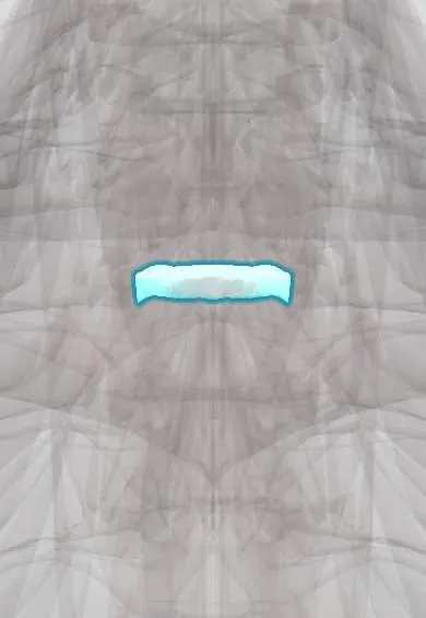
T2–T3 disc !
The T2-T3 disc is the thin cushion between the T2 and T3 vertebrae in the mid-back; it allows the small movement the ribs permit.
Official name: T2-T3
Opens in the Durgical app, no sign-up needed.
What it is
The cushion between T2 and T3: a tough outer ring of fibres (annulus fibrosus) around a soft centre.
What it does
Absorbs load and allows the small amount of movement the rib cage permits.
Key facts
- Where
- Between T2 and T3 bodies; thin thoracic disc; head of rib 3 joins it
- Attaches
- Annulus fibrosus anchored to the endplates and ring apophyses of T2 and T3; anterior and posterior longitudinal ligaments
- Function
- Absorbs and spreads load; herniation rare, often central onto the cord; lateral herniation may irritate the T2 root
Relations
- In front
- Esophagus and trachea, with the longus colli thinning out on the bodies; the aortic arch lies just below and to the left
- Lateral
- Head of rib 3 on the disc and its costovertebral joint; the sympathetic trunk lying on the rib heads
- Behind
- Posterior longitudinal ligament and the spinal cord in a narrow canal
How to find it
Not palpable. Counting spinous processes down from the vertebra prominens (C7), the disc lies roughly level with the tip of the T2 spine, since thoracic spines slope downward, near the superior angle of the scapula.
On imaging
Plain lateral films are blocked by the shoulders at this height (a swimmer's view helps). The level is usually named on sagittal MRI by counting down from C2, as upper thoracic levels are easy to miscount.
Common problems
- Thoracic disc herniations are uncommon; many are found by chance on MRI and cause no symptoms.
- A large one can press on the spinal cord, or cause band-like pain around the chest.
How it is diagnosed
MRI; CT shows whether the herniation is calcified (hardened), which matters for surgery.
Treatment
- Usually physical therapy and pain medicine; surgery only if the spinal cord is pressed.
Clinical notes
- The first two intercostal spaces are fed by the supreme (superior) intercostal artery from the costocervical trunk, not by aortic branches, which matters when segmental vessels are mapped before surgery
- Usually reached from behind and to the side (costotransversectomy): the cord cannot be pushed aside, and the front is crowded by the great vessels
- On the upper chest the C4 skin band borders T2 directly, because C5–T1 have moved out into the arm; a sensory level here is easy to miss unless the arms are tested too
Test yourself
Think of the answer, then tap the question to see it.
Where is pain from a T2 root irritated by a T2–T3 disc felt, and what can it mimic?
In the armpit and inner upper arm (through the intercostobrachial nerve), so it can pass for shoulder or heart pain.
Why is the T2–T3 disc one of the least often herniated discs in the spine?
The ribs and the sternum hold this segment almost still.
Why is the T2–T3 disc usually reached from behind and to the side (costotransversectomy)?
The spinal cord cannot be pushed aside, and the front is crowded by the great vessels.
Educational information, not medical advice. For any symptom, see a health professional.
Its names
- Official Latin (TA2)
- discus intervertebralis T2-T3 TA2 1684
- Official Spanish (DeCS)
- Disco T2-T3 DeCS D007403
- English (TA2)
- T2-T3
- Also called
- fibrous ring · fibrocartilago intervertebralis
Related
Sources: Terminologia Anatomica (TA2); DeCS; Anatomy, Back, Intervertebral Discs; 7.3 The Vertebral Column (OpenStax (Rice University), 2022) · adapted, CC BY 4.0.
Spotted a mistake?We are always improving for the community. Tell us what is wrong and our team will review it.Report a mistake
Representative model, for educational purposes only.