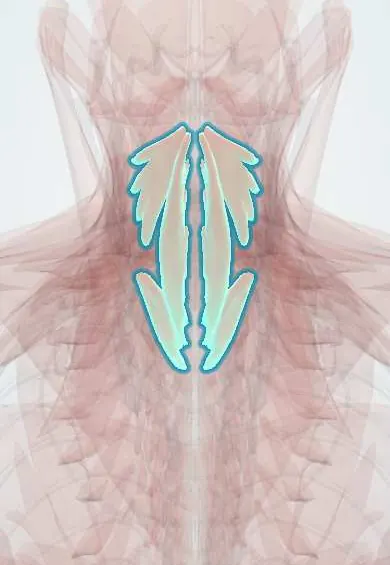
Longus colli !
The longus colli is a long, thin muscle lying on the front of the neck vertebrae, from C1 to T3; it bends the neck forward and steadies it.
Official name: Longus colli muscle
Opens in the Durgical app, no sign-up needed.
What it is
A long, thin muscle lying directly on the front of the neck vertebrae, from C1 down to T3.
What it does
Bends the neck forward and holds it steady, opposing the extensor muscles; it supports the neck's curve.
Key facts
- Origin
- Bodies of C5–T3; transverse processes of C3–C5
- Insertion
- Anterior tubercle of atlas; bodies of C2–C4; transverse processes of C5–C6
- Action
- Flexes the neck; slight rotation and lateral flexion; supports cervical lordosis
- Nerve
- Ventral rami of C2–C6
- Blood supply
- Inferior thyroid, ascending cervical and vertebral arteries
Relations
- In front
- Prevertebral fascia with the sympathetic trunk; further forward the retropharyngeal space, pharynx and oesophagus; the carotid sheath in front and to the side
- Behind
- Vertebral bodies and the anterior longitudinal ligament from C1 to T3
- Medial
- The anterior longitudinal ligament and vertebral bodies, exposed in the midline between the two muscles
- Lateral
- Scalenus anterior and, low in the neck, the vertebral artery between the two muscles; longus capitis above
How to find it
It cannot be felt. In the anterior cervical approach it is the reddish muscle on each side of the midline once the prevertebral fascia is opened; on CT and MRI it lies directly on the front of the vertebral bodies.
Examination
- Craniocervical flexion test: lying on the back with a pressure cuff behind the neck, the person nods in small steps and holds each one; inability to hold the nod, or using sternocleidomastoid instead, points to weak deep neck flexors, common in chronic neck pain and after whiplash
On imaging
Lateral neck radiograph: the prevertebral soft tissue, which includes this muscle, is normally thin (commonly taught upper limits in adults: about 7 mm at C2 and 22 mm at C6); widening suggests haematoma, abscess or tendinitis. CT shows the calcium in front of C1–C2 in calcific tendinitis.
Common problems
- Strained in whiplash.
- In an ACDF it is gently lifted off the front of the spine on each side to place the retractor; the sympathetic trunk lies on it.
How it is diagnosed
Exam; MRI if needed.
Treatment
- Deep neck flexor exercises are a mainstay of neck pain rehabilitation.
Clinical notes
- Acute calcific tendinitis of longus colli: sudden neck pain and painful swallowing with calcium in front of C1–C2 and prevertebral swelling; it mimics a retropharyngeal abscess
- Retractor blades are seated under its medial edges; staying medial to them protects the sympathetic trunk and the vertebral artery
Development
With the other prevertebral muscles it develops from the ventral part of the cervical myotomes, so it is supplied by ventral rami of the cervical spinal nerves, not by dorsal rami like the true back muscles.
Memory aid
Prevertebral soft tissue: 7 at 2, 22 at 6 (millimetres at C2 and C6, adults).
Related conditions
Test yourself
Think of the answer, then tap the question to see it.
What are the extent and action of the longus colli?
It lies on the vertebral bodies from C1 to T3; it flexes the neck and supports the cervical lordosis.
Which structure on the front of the longus colli is at risk if dissection goes lateral to its medial edge?
The cervical sympathetic trunk; injury causes Horner syndrome.
What is acute calcific tendinitis of the longus colli?
Sudden neck pain and painful swallowing with calcium in front of C1–C2 and prevertebral swelling; it mimics a retropharyngeal abscess.
Why is the midline strip between the two longus colli muscles important in anterior cervical surgery?
It is the safe corridor; retractors sit under the muscles' medial edges, protecting the sympathetic trunk and vertebral artery.
Educational information, not medical advice. For any symptom, see a health professional.
Its names
- Official Latin (TA2)
- musculus longus colli TA2 2148
- Official Spanish (DeCS)
- Músculo largo del cuello DeCS D009334
- English (TA2)
- Longus colli muscle
- Also called
- longus cervicis muscle
Related
- C5–C6 disc
- Sympathetic trunk
- Neck nerve roots
- Vertebral artery
- Atlas (C1)
- Axis (C2)
- C3 vertebra
- C4 vertebra
Sources: Terminologia Anatomica (TA2); DeCS; Anatomy, Head and Neck, Prevertebral Muscles; Anatomy of the Human Body (20th ed.): Development of the Muscles (Lea & Febiger (Gray's Anatomy, 1918), 1918) · adapted, Public domain.
Spotted a mistake?We are always improving for the community. Tell us what is wrong and our team will review it.Report a mistake
Representative model, for educational purposes only.