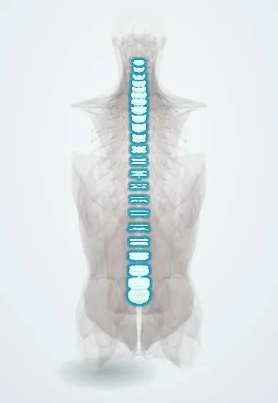
Endplates !
The endplates are thin layers of cartilage and bone capping each vertebral body; they hold the disc and let nutrients reach it.
Official name: Vertebral endplates
Opens in the Durgical app, no sign-up needed.
What it is
Thin layers of cartilage and bone that cap the top and bottom of each vertebral body, against the discs.
What it does
Hold the disc in place and let nutrients pass into the disc, which has almost no blood supply of its own.
Key facts
- Where
- Upper and lower surfaces of each vertebral body against the disc: cartilage endplate over a thin bony endplate, inside the ring apophysis
- Attaches
- Inner annulus fibrosus fibres anchor in the cartilage endplate; outer annulus fibres anchor in the bony ring apophysis
- Function
- Hold the disc to the bone; main route of nutrient diffusion into the avascular disc; weak spot where nucleus herniates into the body (Schmorl node)
Relations
- Superficial
- Disc side: the nucleus pulposus and the inner annulus fibrosus of the adjacent intervertebral disc
- Deep
- Cancellous bone of the vertebral body with its marrow and the capillary loops that feed the disc by diffusion
- Lateral
- The ring apophysis around the rim, and outside it the anterior longitudinal ligament in front and the posterior longitudinal ligament behind
On imaging
MRI shows the endplate as a dark line and Modic changes in the marrow beside it. Radiographs and CT show Schmorl nodes as rounded defects with a sclerotic rim. In spondylodiscitis the endplates on both sides of a disc are eroded and the disc is bright on T2, which helps tell infection from Modic type 1.
Common problems
- Small cracks or bone-marrow changes next to a worn disc (Modic changes) that can be linked to back pain.
- Schmorl's nodes: the disc pushes into the vertebral body through the endplate.
How it is diagnosed
MRI shows endplate changes; X-ray or CT shows Schmorl's nodes.
Treatment
- Usually treated like back pain in general. In a fusion, the surgeon scrapes the disc off the endplates so the bone graft can grow across.
Clinical notes
- Spondylodiscitis in adults usually seeds the vascular bone next to the endplate and spreads across the disc to the next body
- Osteoporotic compression fractures usually begin as endplate fractures, often of the upper endplate
- Modic type 1 changes have been associated with low back pain more often than other types, although the link is debated
Anatomical variants
- Cupid's bow contour: a smooth, paired concavity of the lower endplates of the lower lumbar vertebrae, seen on frontal films and CT, is a normal variant and is not a fracture
Development
The ring apophysis ossifies from secondary centres around puberty and fuses with the body in early adulthood; before fusion the nucleus can herniate under it and lift off a small triangular corner (limbus vertebra), which can mimic a fracture.
Memory aid
Modic 1-2-3: wet (oedema), fat, hard (sclerosis).
Related conditions
Test yourself
Think of the answer, then tap the question to see it.
Under axial compression, which usually fails first: the vertebral endplate or the annulus fibrosus?
The endplate usually cracks first; it is the weakest link of the vertebra–disc unit, and osteoporotic compression fractures often begin there.
How do Modic types 1, 2 and 3 of the vertebral endplates look on MRI?
Type 1 oedema (dark T1, bright T2), type 2 fatty marrow (bright T1 and T2), type 3 sclerosis (dark on both).
What is a Schmorl node of the vertebral endplate?
Herniation of the nucleus pulposus through the endplate into the vertebral body, seen as a rounded defect with a sclerotic rim.
Why is calcification of the cartilage endplate with age linked to disc degeneration?
The endplate is the main route of nutrient diffusion into the avascular disc; calcification and falling capillary supply cut that diffusion.
Educational information, not medical advice. For any symptom, see a health professional.
Its names
- Official Latin (TA2)
- facies intervertebralis TA2 1013
- Official Spanish (DeCS)
- Platillos vertebrales
- English (TA2)
- Vertebral endplates
Related
Sources: Terminologia Anatomica (TA2); Anatomy, Back, Intervertebral Discs.
Spotted a mistake?We are always improving for the community. Tell us what is wrong and our team will review it.Report a mistake
Representative model, for educational purposes only.