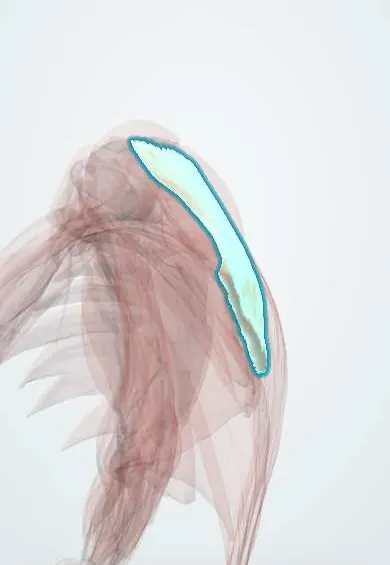
Middle trapezius !
The middle trapezius runs across the upper back from the spine to the shoulder blade; it pulls the shoulder blades back together.
Official name: Trapezius, middle (transverse) part
Opens in the Durgical app, no sign-up needed.
What it is
The middle part of the trapezius, between the spine and the shoulder blade.
What it does
Pulls the shoulder blades back together.
Key facts
- Origin
- Spinous processes C7–T3
- Insertion
- Acromion and spine of the scapula
- Action
- Retracts the scapula
- Nerve
- Accessory nerve (CN XI)
- Blood supply
- Transverse cervical artery
Relations
- Superficial
- Skin and fascia of the upper back between the shoulder blades
- Deep
- Rhomboid minor and rhomboid major medially, with the dorsal scapular nerve and artery deep to them; supraspinatus near its insertion
- Medial
- Spinous processes C7–T3 and the supraspinous ligament; with the other side it forms a tendinous diamond around C7
- Lateral
- Acromion and spine of the scapula
How to find it
Lying prone with the arm abducted to 90° and turned out (thumb up), lifting the arm toward the ceiling squeezes the shoulder blades together and the horizontal band between the spine and the scapula tightens under the fingers.
Examination
- Manual muscle test (prone, arm at 90° abduction, examiner pressing the arm toward the floor): weakness with the scapula drifting away from the spine points to CN XI injury or marked deconditioning
- Winging pattern: winging that grows with resisted abduction and eases on forward flexion points to the trapezius (accessory nerve); winging on a wall push-up points to the serratus anterior (long thoracic nerve)
On imaging
On axial CT or MRI of the upper chest it is the most superficial muscle between the spine and the scapula, over the rhomboids. Accessory nerve palsy shows as atrophy and fatty change. Scapular winging is a clinical sign; imaging is used mainly to compare sides and to rule out a mass. Ultrasound measures its thickness at the bedside.
Clinical notes
- After accessory nerve injury the scapula sits lower and further from the spine at rest, and the shoulder may ache from traction on the brachial plexus
- Weak middle and lower fibres with a protracted, forward-tipped scapula is a common picture in scapular dyskinesis and in overhead athletes with shoulder pain
Related conditions
Test yourself
Think of the answer, then tap the question to see it.
Which muscles are the main retractors of the scapula?
The middle trapezius, with the rhomboid major and rhomboid minor.
How does trapezius winging differ from serratus anterior winging?
Trapezius (accessory nerve): lateral winging, the scapula slides down and away, worse on abduction. Serratus anterior: medial winging, worse on pushing forward.
Where does the middle trapezius insert?
On the acromion and the upper lip of the spine of the scapula, facing the origin of the rear deltoid on the lower lip.
Educational information, not medical advice. For any symptom, see a health professional.
Its names
- Official Latin (TA2)
- pars transversa musculi trapezii TA2 2228
- Official Spanish (DeCS)
- Porción transversa del músculo trapecio
- English (TA2)
- Trapezius, middle (transverse) part
Related
Sources: Terminologia Anatomica (TA2); Anatomy, Back, Trapezius.
Spotted a mistake?We are always improving for the community. Tell us what is wrong and our team will review it.Report a mistake
Representative model, for educational purposes only.